Dermatología en Costa Rica

Tuesday, October 31, 2023

Edgar Allan Poe on daydreaming

Wednesday, October 25, 2023

PeerView - Bridging the Gap to Increased Patient Satisfaction in Moderate to Severe Atopic Dermatitis: Understanding the Role and Clinical Utility of Targeted Therapy

PVI, PeerView Institute for Medical Education, respects and appreciates your opinions. To obtain CME/CE/AAPA credit, please complete the post-test. To assist us in evaluating the effectiveness of this activity and to make recommendations for future educational offerings, please take a few minutes to complete the evaluation form.

You passed the post-test! Please review your answers below and tap Continue at the bottom of the page to submit.

Question 1/5
Which of the following cytokines is a driver of chronic type 2 inflammation in AD, binds directly to sensory nerves, increases sensation of itch, and is inhibited by lebrikizumab?
  • IL-13
Question 2/5
Your patient is a 19-year-old Black man who claims the topical corticosteroids he has been using for AD are not working. He complains that at least once per month, persistent itching keeps him up almost all night, causing him to scratch to the point of bleeding. Upon examination, you observe rashes that appear violet in color, significant lichenification, oozing lesions, some patches of dyspigmentation, and signs of a secondary bacterial infection from scratching on his torso, arms, and legs. How would you categorize the severity of his AD?
  • Moderate to severe
Question 3/5
Consider the above 19-year-old male patient. What therapies would you consider?
  • Suggest a switch to a topical calcineurin inhibitor and add a biologic therapy
Question 4/5
Which of the following statements is true regarding the use of topical therapies, with or without systemic therapy, for AD?
  • A substantial number of adult and adolescent patients, and their physicians, report dissatisfaction with topical AD therapies, with or without systemic therapy
Question 5/5
You are evaluating a patient with moderate to severe AD who has not experienced relief after months on topical therapies and wants to know what they can do next. Your patient has also expressed a desire to not have to remember to take pills on a daily basis or to have to come to your office frequently. What would your next step be?
  • Suggest they try a biologic therapy such as dupilumab or tralokinumab (or lebrikizumab, if approved)

Sent from my iPhone

Benjamin Hidalgo-Matlock
Skin Care Physicians of Costa Rica

Clinica Victoria en San Pedro: 4000-1054
Momentum Escazu: 2101-9574

Please excuse the shortness of this message, as it has been sent from
a mobile device.

TATN

A PERMANENT ADDITION TO THE LIST OF TRANSIENT NEONATAL DERMATOSES: TRANSIENT ABDOMINAL TELANGIECTASIA OF THE NEWBORN


By Warren R. Heymann, MD, FAAD
Oct. 25, 2023
Vol. 5, No. 42

Any parent looking at their newborn child will wonder if the skin lesions they observe are worrisome. During the neonatal period, defined as the first four weeks of life, most cutaneous findings are transient and benign; occasionally, they may be manifestations of serious disease. 

Transient vascular changes include cutis marmorata (CM, a reticulated mottling of the skin observed symmetrically on the trunk and extremities, worsened by cold and resolving with warmth) and the harlequin color change (HCC, occurring when the neonate lies on their side, with erythema on the dependent side and blanching on the contralateral side, lasting for 30 seconds to 20 minutes). CM may last a few months, while HCC resolves in a few weeks. Both disorders are considered physiologic; although HCC has been attributed to hypothalamic immaturity causing sympathetic autonomic dysfunction affecting capillary bed tonus. (1,2)

Benign, transient disorders in the neonatal period include milia, miliaria, transient pigmentary lines, erythema toxicum neonatorum, transient neonatal pustular melanosis, and benign cephalic pustulosis. (Infectious neonatal pustular dermatoses due to Staphylococcus, Candida, Herpes virus, or other microbes must be ruled out, if clinically warranted). (1,3,4)

Other transient dermatoses of the newborn reflect underlying pathology. Transient bullous dermolysis of the newborn (TBDN) is a rare variant of dystrophic epidermolysis bullosa caused by autosomal dominant or recessive mutations in COL7A1. TBDN is characterized by subepidermal blistering at birth or shortly thereafter, followed by rapid improvement with minimal scarring or pigmentation. (5) According to Brazzelli et al. "Transient myeloproliferative disorder (TMD) is a spontaneously resolving clonal myeloid proliferation characterized by circulating megakaryoblasts in the peripheral blood that is restricted to neonates with Down syndrome (DS) or those with trisomy 21 mosaicism. Cutaneous manifestations of TMD are observed in only 5% of affected neonates and present as a diffuse eruption of erythematous, crusted papules, papulovesicles, and pustules, often with prominent and initial facial involvement." TMD resolves spontaneously in most cases, although there is a low potential to develop acute myeloid leukemia. (6)

Image with permission from Dr. Cécile Juzot.

In a retrospective, observational, multicenter study, Juzot et al reported 20 newborns who developed, at a median age of 7 days, large abdominal patches of radially arranged purplish telangiectasia in a bilateral and symmetrical pattern relative to the midline, creating a "butterfly wing" pattern, labeling this entity "transient abdominal telangiectasia of the newborn" (TATN). Clinical examination was normal in 13 newborns; 6 newborns had abdominal distention, and one newborn had poor weight gain due to inadequate breastfeeding. Dermoscopy confirmed telangiectasis; no biopsies were performed. In 15 of the 20 newborns, the umbilical stump had already detached by the time the lesions appeared. Fourteen newborns underwent abdominal ultrasonography, which was unremarkable for 10 of the children, 2 of whom had infrequent stools. Of the 4 scans showing abnormalities, findings included incidental bilateral ovarian cysts, a pelvic mass (in a newborn with rhabdomyosarcoma), stercoral stasis (in a child with Hirschsprung disease), and poorly visualized mesenteric vessels (in the context of abdominal distention). Most lesions spontaneously resolved within 3 months and did not reoccur for 19 newborns. Although the etiology is unknown, the working hypothesis is that increased intraabdominal pressure affects the cutaneous microcirculation. The authors conclude, "While most cases of TATN regress spontaneously and are benign, their presence should lead the clinician to check the medical history, perform a careful clinical abdominal examination, and where appropriate consider abdominal ultrasound to rule out a pelvic or intraabdominal mass. Larger case studies are required to further characterize this entity. The mechanism leading to the manifestation of TATN still remains to be elucidated."

Point to Remember: Pediatric dermatologists must carefully assess any neonate with dermatologic findings. Many are benign and transient. A new neonatal entity has been described — transient abdominal telangiectasia of the newborn (TANT). TANT is a benign disorder. Ultrasound imaging to rule out intrabdominal pathology is indicated if abdominal distention is observed. 

Our expert's viewpoint

Cecile Juzot, MD
Department of Dermatology, Centre Hospitalier Universitaire de Nantes, France

Since our work was published in 2021, I have been able to see a new case of transient abdominal telangiectasia of the newborn (TATN). It was a girl, born at full term. The pregnancy had been marked by lysis of a twin in the first trimester. We examined her at 3 days of age for a hypochromic macule, sometimes purplish, on the left buttock with a similar lesion on the left lower limb, unnoticed on the day of birth. Dermatological and general examination was otherwise normal. On the 19th day of life, I noticed TATN and a discreetly distended abdomen. The telangiectasias reappeared until about 2 months of age, during episodes of abdominal distention. Abdominal ultrasound was normal. The vascular lesion of the left lower limb seems to be related to a cutis marmorata telangiectatica congenita and further explorations are ongoing. Other cases have been described. Cutrone et al described 4 cases, 3 girls and 1 boy, including a premature one, all in the context of abdominal distension. (8) Bosma et alrecently published the case of a boy. (9)

These new cases seem to confirm that TATN are frequent, probably underestimated, frequently occur in a context of abdominal distension, and rapidly disappear spontaneously.

  1. O'Connor NR, McLaughlin MR, Ham P. Newborn skin: Part I. Common rashes. Am Fam Physician. 2008 Jan 1;77(1):47-52. PMID: 18236822.

  2. Tsuboi K, Tsuboi N, Nosaka N, Nishimura N, Nakagawa S. Neonatal Harlequin color change associated with Prostaglandin E1 administration. Pediatr Int. 2021 May;63(5):610-611. doi: 10.1111/ped.14581. PMID: 34002472.

  3. Terry M, Marlowe Stewart D. Transient Pigmentary Lines of the Newborn in a Postmortem Examination: A Case Report. Am J Forensic Med Pathol. 2019 Jun;40(2):171-174. doi: 10.1097/PAF.0000000000000461. PMID: 30689604.

  4. Reginatto FP, Villa DD, Cestari TF. Benign skin disease with pustules in the newborn. An Bras Dermatol. 2016 Apr;91(2):124-34. doi: 10.1590/abd1806-4841.20164285. PMID: 27192509; PMCID: PMC4861557.

  5. Shi BJ, Zhu XJ, Liu Y, Hao J, Yan GF, Wang SP, Wang XY, Diao QC. Transient bullous dermolysis of the newborn: a novel de novo mutation in the COL7A1 gene. Int J Dermatol. 2015 Apr;54(4):438-42. doi: 10.1111/ijd.12704. PMID: 25800346.

  6. Brazzelli V, Segal A, Bernacca C, Tchich A, Bolcato V, Croci G, Mina T, Zecca M, Zanette S, Stronati M. Neonatal vesiculopustular eruption in Down syndrome and transient myeloproliferative disorder: A case report and review of the literature. Pediatr Dermatol. 2019 Sep;36(5):702-706. doi: 10.1111/pde.13931. Epub 2019 Jul 29. PMID: 31355466.

  7. Juzot C, Aubert H, Bessis D, Boccara O, Bourrat E, Chiaverini C, Flamant C, Fournet M, Hubiche T, Labrèze C, Martin L, Piram M, Seta V, Finon A, Maruani A, Barbarot S; "Groupe de Recherche Clinique de Dermatologie Pédiatrique". Transient abdominal telangiectasia of the newborn. Pediatr Dermatol. 2021 Jul;38(4):864-867. doi: 10.1111/pde.14620. Epub 2021 Jun 21. PMID: 34152036.

  8. Cutrone M, Van Gysel D. Transient abdominal telangiectasia of the newborn: Four new cases with abdominal distension. Pediatr Dermatol. 2023 May 31. doi: 10.1111/pde.15351. Epub ahead of print. PMID: 37258091. 

  9. Bosma AL, Van der Zwaan D. Een pasgeborene met vaattekening op de buik [Abdominal telangiectasias in a newborn]. Ned Tijdschr Geneeskd. 2022 Oct 5;166:D6960. Dutch. PMID: 36300458.


All content found on Dermatology World Insights and Inquiries, including: text, images, video, audio, or other formats, were created for informational purposes only. The content represents the opinions of the authors and should not be interpreted as the official AAD position on any topic addressed. It is not intended to be a substitute for professional medical advice, diagnosis, or treatment.

DW Insights and Inquiries archive

Explore hundreds of Dermatology World Insights and Inquiries articles by clinical area, specific condition, or medical journal source.


Sent from my iPhone

Benjamin Hidalgo-Matlock
Skin Care Physicians of Costa Rica

Clinica Victoria en San Pedro: 4000-1054
Momentum Escazu: 2101-9574

Please excuse the shortness of this message, as it has been sent from
a mobile device.

Sunday, October 22, 2023

Jaks y acne…

Review Estimates Acne Risk With JAK Inhibitor Therapy

Heidi Splete

October 19, 2023

TOPLINE: 

Use of Janus kinase (JAK) inhibitors is associated with a nearly fourfold increase in risk of acne compared with placebo, according to an analysis of 25 JAK inhibitor studies.

METHODOLOGY: 

  • Acne has been reported to be an adverse effect of JAK inhibitors, but not much is known about how common acne is overall and how incidence differs between different JAK inhibitors and the disease being treated.

  • For the systematic review and meta-analysis, researchers identified 25 phase 2 or 3 randomized, controlled trials that reported acne as an adverse event associated with the use of JAK inhibitors.

  • The study population included 10,839 participants (54% male, 46% female).

  • The primary outcome was the incidence of acne following a period of JAK inhibitor use.

TAKEAWAY: 

  • Overall, the risk of acne was significantly higher among those treated with JAK inhibitors in comparison with patients given placebo in a pooled analysis (odds ratio [OR], 3.83).

  • The risk of acne was highest with abrocitinib (OR, 13.47), followed by baricitinib (OR, 4.96), upadacitinib (OR, 4.79), deuruxolitinib (OR, 3.30), and deucravacitinib (OR, 2.64). By JAK inhibitor class, results were as follows: JAK1-specific inhibitors (OR, 4.69), combined JAK1 and JAK2 inhibitors (OR, 3.43), and tyrosine kinase 2 inhibitors (OR, 2.64).

  • In a subgroup analysis, risk of acne was higher among patients using JAK inhibitors for dermatologic conditions in comparison with those using JAK inhibitors for nondermatologic conditions (OR, 4.67 vs 1.18).

  • Age and gender had no apparent impact on the effect of JAK inhibitor use on acne risk.

IN PRACTICE: 

"The occurrence of acne following treatment with certain classes of JAK inhibitors is of potential concern,as this adverse effect may jeopardize treatment adherence among some patients," the researchers wrote. More studies are needed "to characterize the underlying mechanism of acne with JAK inhibitor use and to identify best practices for treatment," they added.

SOURCE: 

The lead author was Jeremy Martinez, MPH, of Harvard University, Boston. The study was published online in JAMA Dermatology on October 18, 2023.

LIMITATIONS: 

The review was limited by the variable classification and reporting of acne across studies, the potential exclusion of relevant studies, and the small number of studies for certain drugs.

DISCLOSURES: 

The studies were mainly funded by the pharmaceutical industry. Martinez disclosed no relevant financial relationships. Several co-authors have relationships with multiple companies, including Dexcel Pharma Technologies, AbbVie, Concert, Pfizer, 3Derm Systems, Incyte, Aclaris, Eli Lilly, Concert, Equillium, ASLAN, ACOM, and Boehringer Ingelheim.

For more news, follow Medscape on FacebookXInstagram, and YouTube.


    Sent from my iPhone

    Benjamin Hidalgo-Matlock
    Skin Care Physicians of Costa Rica

    Clinica Victoria en San Pedro: 4000-1054
    Momentum Escazu: 2101-9574

    Please excuse the shortness of this message, as it has been sent from
    a mobile device.

    Tuesday, October 10, 2023

    Spironolactone for HS

    Spironolactone Safe, Effective Option for Women With Hidradenitis Suppurativa

    Doug Brunk

    October 04, 2023

    CARLSBAD, CALIF. – Spironolactone may be an effective and safe treatment option for women with hidradenitis suppurativa (HS), regardless of whether they report having menstrual HS flares or have been diagnosed with polycystic ovary syndrome (PCOS).

    Those are the key findings from a single-center retrospective study that Jennifer L. Hsiao, MD, and colleagues presented during a poster session at the annual symposium of the California Society of Dermatology & Dermatologic Surgery.

    In an interview after the meeting, Dr. Hsiao, a dermatologist who directs the hidradenitis suppurativa clinic at the University of Southern California, Los Angeles, said that hormones are thought to play a role in HS pathogenesis given the typical HS symptom onset around puberty and fluctuations in disease activity with menses (typically premenstrual flares) and pregnancy. "Spironolactone, an anti-androgenic agent, is used to treat HS in women; however, there is a paucity of data on the efficacy of spironolactone for HS and whether certain patient characteristics may influence treatment response," she told this news organization. "This study is unique in that we contribute to existing literature regarding spironolactone efficacy in HS and we also investigate whether the presence of menstrual HS flares or polycystic ovarian syndromeinfluences the likelihood of response to spironolactone."

    For the analysis, Dr. Hsiao and colleagues retrospectively reviewed the medical records of 53 adult women with HS who were prescribed spironolactone and who received care at USC's HS clinic between January 2015 and December 2021. They collected data on demographics, comorbidities, HS medications, treatment response at 3 and 6 months, as well as adverse events. They also evaluated physician-assessed response to treatment when available.

    The mean age of patients was 31 years, 37% were White, 30.4% were Black, 21.7% were Hispanic, 6.5% were Asian, and the remainder were biracial. The mean age at HS diagnosis was 25.1 years and the three most common comorbidities were acne (50.9%), obesity(45.3%), and anemia (37.7%). As for menstrual history, 56.6% had perimenstrual HS flares and 37.7% had irregular menstrual cycles. The top three classes of concomitant medications were antibiotics (58.5%), oral contraceptives (50.9%), and other birth controlmethods (18.9%).

    The mean spironolactone dose was 104 mg/day; 84.1% of the women experienced improvement of HS 3 months after starting the drug, while 81.8% had improvement of their HS 6 months after starting the drug. The researchers also found that 56.6% of women had documented perimenstrual HS flares and 7.5% had PCOS.

    "Spironolactone is often thought of as a helpful medication to consider if a patient reports having HS flares around menses or features of PCOS," Dr. Hsiao said. However, she added, "our study found that there was no statistically significant difference in the response to spironolactone based on the presence of premenstrual flares or concomitant PCOS." She said that spironolactone may be used as an adjunct therapeutic option in patients with more severe disease in addition to other medical and surgical therapies for HS. "Combining different treatment options that target different pathophysiologic factors is usually required to achieve adequate disease control in HS," she said.

    Dr. Hsiao acknowledged certain limitations of the study, including its single-center design and small sample size. "A confounding variable is that some patients were on other medications in addition to spironolactone, which may have influenced treatment outcomes," she noted. "Larger prospective studies are needed to identify optimal dosing for spironolactone therapy in HS as well as predictors of treatment response."

    Adam Friedman, MD, professor and chair of dermatology at George Washington University, Washington, who was asked to comment on the study, said that with only one FDA-approved systemic medication for the management of HS (adalimumab), "we off-label bandits must be creative to curtail the incredibly painful impact this chronic, destructive inflammatory disease can have on our patients."

    "The evidence supporting our approaches, whether it be antibiotics, immunomodulators, or in this case, antihormonal therapies, is limited, so more data is always welcome," said Dr. Friedman, who was not involved with the study. "One very interesting point raised by the authors, one I share with my trainees frequently from my own experience, is that regardless of menstrual cycle abnormalities, spironolactone can be impactful. This is important to remember, in that overt signs of hormonal influences is not a requisite for the use or effectiveness of antihormonal therapy."

    Dr. Hsiao disclosed that she is a member of board of directors for the Hidradenitis Suppurativa Foundation. She has also served as a consultant for AbbVie, Aclaris, Boehringer Ingelheim, Novartis, UCB, as a speaker for AbbVie, and as an investigator for Amgen, Boehringer Ingelheim, and Incyte. Dr. Friedman reported having no relevant financial disclosures.

    This article originally appeared on MDedge.com, part of the Medscape Professional Network.


      Sent from my iPhone

      Benjamin Hidalgo-Matlock
      Skin Care Physicians of Costa Rica

      Clinica Victoria en San Pedro: 4000-1054
      Momentum Escazu: 2101-9574

      Please excuse the shortness of this message, as it has been sent from
      a mobile device.

      Eczema types: Stasis dermatitis self-care

      Eczema types: Stasis dermatitis self-care

      Eczema types: Stasis dermatitis self-care

      12 healthy habits that help prevent worsening stasis dermatitis

      If you have stasis dermatitis, a treatment plan along with self-care can get the disease under control and prevent it from worsening. Here are the healthy habits that dermatologists recommend for their patients who have stasis dermatitis.

      1. Elevate your legs above the heart: Do this throughout the day. If possible, dermatologists recommend that you elevate your legs above your heart:

        • Once every 2 hours for 15 minutes
        • While you sleep (keep your legs elevated with pillows)

        Why elevating your legs helps

        When you raise your legs above your heart, you improve blood flow.

        Woman elevating legs to reduce swelling
      2. Take breaks when you must sit or stand for an hour or longer. If you must sit or stand for long periods, take a break every hour and walk briskly for 10 minutes. This will jump-start your circulation.

      3. Get physical. Exercise can improve your circulation and strengthen your calf muscles. Walking is an especially good exercise for people who have stasis dermatitis. Be sure to build up slowly and ask your dermatologist how often you should exercise.

      4. Wear loose-fitting cotton clothing. Wool and other rough fabrics, polyester, and rayon can irritate skin with stasis dermatitis and lead to a flare-up.

        A loose fit is also important. Tight waistbands and snug pants interfere with your circulation. If clothing rubs against stasis dermatitis, the fabric can irritate the sensitive skin.

      5. Use your compression garment if your dermatologist recommends one. Compression can:

        • Improve the circulation in your legs
        • Prevent open sores
        • Reduce your risk of another flare
        If you have trouble putting on your compression garment or wearing it causes discomfort, tell your dermatologist. This is a common problem. Your dermatologist can provide some helpful tips or write a prescription for physical therapy.

        A physical therapist can offer tips for reducing the pain when you put on the garment. Most patients find that once they start wearing the compression garment, their swelling decreases within a few weeks. With less swelling, they start to feel better.
      6. Avoid injuring the area and aggravating the stasis dermatitis. The skin with stasis dermatitis is very sensitive. If you injure or aggravate the area, it could lead to an infection or open sores.

        To avoid irritating the skin with stasis dermatitis, avoid touching anything that could irritate it, such as:

        • Pet hair
        • Plants
        • Grass
        • Cleaning products
        • Perfume
        • Skin care products that contain fragrance (use only products labeled "fragrance-free.")

        Scratching can aggravate stasis dermatitis, which could lead to an infection

        To help patients avoid scratching, dermatologists offer these tips:

        • Apply your medication as directed
        • Place a clean, cool compress on the itchy area for 15 minutes
        • Slather on a fragrance-free moisturizer like petroleum jelly
        • Take a cool bath in colloidal oatmeal

        If you injure your leg or irritate the skin, contact your dermatologist.
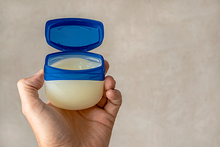
        The hand of a male person holds a container with petroleum jelly
      7. Moisturize dry skin. Moisturizer helps prevent scaly skin and irritation. Petroleum jelly works well for most patients. If you prefer to use another moisturizer, choose an ointment or thick cream that says "fragrance-free" on the container.

      8. Take care when bathing. Soaps and rough-textured towels or bath sponges can irritate skin with stasis dermatitis. Dermatologists recommend the following to their patients with stasis dermatitis:

        • Use a mild, fragrance-free cleanser rather than soap. When you shower or take a bath, use only this cleanser. Rinsing soap from other parts of your body can cause the soap to run down your body, which can irritate skin with stasis dermatitis.
        • After bathing, gently pat the water from your skin with a clean towel. You'll want to keep a bit of water on the skin with stasis dermatitis.
        • Within 2 minutes of bathing, apply petroleum jelly or a thick, creamy moisturizer that is fragrance-free on your damp skin. This helps to keep moisture in your skin. Keeping your skin moisturized helps to prevent scaly skin and irritation.
      9. Reach and stay at a healthy weight. Staying at a healthy weight can reduce swelling and improve your overall health.

      10. Drink 8 glasses of water every day. This can improve circulation and reduce swelling.

      11. Limit salt. Too much salt can decrease your blood flow. Even if you never salt your food, you may be getting too much salt. According to the American Heart Association, the average American consumes 3,400 milligrams of sodium every day. The recommended daily amount is 1,500 milligrams or less.3

        For tips that can help you cut back on the salt, go to, How to reduce sodium.

      12. Keep your dermatology appointments. Stasis dermatitis is a condition that you may have for life. Learning how to manage it and finding out what works best for you can take time. The time spent learning what to do will pay off. Most patients find that once they know what to do, they can manage the disease at home with healthy habits and medication as needed to treat flare-ups.

        If you need a dermatologist, you can find one by going to, Find a dermatologist.

      Related AAD resources


      Images
      Getty Images

      References
      American Academy of Dermatology. "Stasis dermatitis and leg ulcers." Basic Dermatology Curriculum. Last accessed August 28, 2020.

      3 American Heart Association. "9 out of 10 Americans eat too much sodium." Page last accessed August 31, 2020.

      Flugman SL, Clark RA. [editor: Elston DM] "Stasis dermatitis." Medscape. Last updated Mar 27, 2020.

      Nedorost S, White S, et al. "Development and implementation of an order set to improve value of care for patients with severe stasis dermatitis." J Am Acad Dermatol. 2019 Mar;80(3):815-7.

      Reider N, Fritsch PO. "Other eczematous eruptions." In: Bolognia JL, et al. Dermatology. (fourth edition). Mosby Elsevier, China, 2018:235-6.

      Sundaresan S, Migden MR, et al. "Stasis dermatitis: Pathophysiology, evaluation, and management. Am J Clin Dermatol. 2017;18(3):383-90.


      Sent from my iPhone

      Benjamin Hidalgo-Matlock
      Skin Care Physicians of Costa Rica

      Clinica Victoria en San Pedro: 4000-1054
      Momentum Escazu: 2101-9574

      Please excuse the shortness of this message, as it has been sent from
      a mobile device.