Dermatología en Costa Rica

Friday, April 27, 2018

Lamotrigeno y reaccion inmune

FDA: Lamotrigine Can Cause Serious Immune Reaction

By Amy Orciari Herman

Edited by André Sofair, MD, MPH, and William E. Chavey, MD, MS

Lamotrigine, used to treat seizures and bipolar disorder, can cause hemophagocytic lymphohistiocytosis (HLH), the FDA has warned. HLH is a potentially fatal, systemic immune reaction.

From 1994 through 2017, there have been eight suspected or confirmed cases of HLH linked to lamotrigine. All occurred within 24 days of starting treatment. One patient died.

HLH usually starts with a persistent fever. Patients must meet five of the following eight criteria for an HLH diagnosis:

  • Fever and rash
  • Enlarged spleen
  • Cytopenias
  • High triglyceride or low fibrinogen levels
  • High ferritin levels
  • Hemophagocytosis detected via biopsy of bone marrow, the spleen, or lymph nodes
  • Reduced natural killer (NK) cell activity
  • Increased blood levels of CD25

Early diagnosis and treatment are critical. Accordingly, the FDA advises clinicians to promptly evaluate lamotrigine recipients who develop fever or rash, and to stop treatment if they suspect HLH or another immune-related reaction.

FDA MedWatch safety alert (Free)

Background: NEJM Journal Watch Neurology coverage of potential association between lamotrigine and meningitis (Your NEJM Journal Watch registration required)


Benjamin Hidalgo-Matlock
Skin Care Physicians of Costa Rica
4000-1054
2101-9574
Please excuse the shortness of this message, as it has been sent from a mobile device.

Thursday, April 26, 2018

Uso de SPF mayor de 50?

Original article
SPF 100+ sunscreen is more protective against sunburn than SPF 50+ in actual use: Results of a randomized, double-blind, split-face, natural sunlight exposure clinical trial

Referred to by
Kenneth G. Linden
Commentary: Sunscreen sun protection factor (SPF)
Journal of the American Academy of Dermatology, Volume 78, Issue 5, May 2018, Pages 911-912
Purchase PDF

Background

The value of additional photoprotection provided by use of high–sun protection factor (SPF) sunscreens is controversial, and limited clinical evidence exists.

Objective

To compare the sunburn protection provided by SPF 100+ and SPF 50+ sunscreen in conditions of actual use.

Methods

A total of 199 healthy men and women (≥18 years) participated in a natural sunlight, single-exposure, split-face, randomized, double-blind study in Vail, Colorado. Each participant wore both sunscreens simultaneously during activities, with no use restrictions other than designation of the treatment area. Erythema was clinically assessed on the day following exposure. Comparative efficacy was evaluated through bilateral comparison of sunburn between treatment areas and erythema score, as evaluated separately for each treatment area.

Results

Following an average 6.1 ± 1.3 hours of sun exposure, investigator-blinded evaluation identified 55.3% of the participants (110 of 199) as more sunburned on the SPF 50+ protected side and 5% (10 of 199) on the SPF 100+ protected side. After exposure, 40.7% of the participants (81 of 199) exhibited increased erythema scores (by ≥1) on the SPF 50+ protected side as compared with 13.6% (27 of 199) on the SPF 100+ protected side.

Limitations

Single-day exposure may not extrapolate to benefits of longer-term protection.

Conclusion

SPF 100+ sunscreen was significantly more effective in protecting against sunburn than SPF 50+ sunscreen in actual use conditions.

Tuesday, April 24, 2018

caida de cabello por bloqueadores de estrogenos.

Novel Study of Endocrine Therapy-Induced Hair Loss

Pam Harrison

April 17, 2018

It's not only chemotherapy that causes breast cancer patients to lose their hair. Treatment with endocrine therapy (ie, tamoxifen or an aromatase inhibitor) can also cause hair loss and have a negative impact on quality of life, even when the loss is only mild in severity, a novel study suggests.

On the other hand, treatment with topical minoxidil (Rogaine, Johnson & Johnson) can restore hair growth, so the loss is potentially treatable, the investigators add.

"There are tens of thousands of breast cancer survivors on endocrine therapies, and the remarkable improvements in patient survival that have been achieved through their care have led to patients now being concerned about quality of life issues, like partial hair loss," senior author Mario Lacouture, MD, Memorial Sloan Kettering Cancer Center (MSKCC), New York City, told Medscape Medical News in an email.

The study is the first to characterize endocrine therapy–induced alopecia (EIA) in patients with breast cancer. EIA has been anecdotally reported but never systematically described. The study was published online April 11 in JAMA Dermatology.

The investigators retrospectively identified a cohort of 112 female patients seen at MSKCC, all of whom had EIA.

"Those who either previously received cytotoxic chemotherapy or had a previous diagnosis of alopecia or any scalp condition were excluded from the study," the investigators note.

Standardized clinical photographs of the scalp were taken from several angles with the hair parted and combed in the center. Responses to a questionnaire concerning hair were collected in order to assess the effect that alopecia had on patients' quality of life. At baseline, the prevailing trichoscopic feature was the presence of vellus hairs as well as intermediate- and thick-density terminal hair shafts, the researchers report.

Patients were treated with topical minoxidil 5%. The effects of treatment were measured by a single investigator at 3 and 6 months.

The researchers note that vellus hairs may be present as a result of unsynchronized miniaturization of hair follicles, one of the hallmarks of so-called androgenetic alopecia. As Lacouture explained, androgenetic alopecia is a pattern of hair loss similar to that which occurs in men and women with age. It is characterized by a thinning of hair on the temples, a receding hairline, and loss of hair at the top of the head.

In the current study, evaluation of the standardized clinical photographs, available for 93% of the group, showed that there was a more prominent recession of hair in the frontotemporal area in 76% of patients than in the midanterior hairline, the investigators note.

"Also, the specific type of alopecia seen in 86 patients (83%) was mild to moderate alopecia on the crown area of the scalp," they add.

Moreover, 92% of patients developed grade 1 alopecia; only 8% of the group developed grade 2 alopecia. Alopecia severity was graded using the Common Terminology Criteria for Adverse Events scale.

Data on alopecia-related quality of life were available for slightly fewer than half of the group (46%). For those patients, the mean score on the hair questionnaire was 25.6.

This score includes measures of emotions, functioning, symptoms, stigmatization, and self-confidence. The highest negative score on the hair questionnaire was in the emotional domain (P < .001).

Grade 1 alopecia represents hair loss of less than 50% that does not require camouflage.

Patients appeared to be negatively affected by even grade 1 hair loss, suggesting, Lacouture indicated, that "not only complete hair loss as seen with chemotherapy is affecting women but also hair thinning or partial hair loss will result in a negative psychosocial impact."

Minoxidil Response

The investigators were able to assess response to topical minoxidil 5% in 41% of the group. Topical minoxidil 2% and 5% is the only agent approved by the US Food and Drug Administration for the treatment of androgenetic alopecia in women.

The investigators determined that 80% of patients who were treated with topical minoxidil experienced either moderate to significant improvement in alopecia.

The researchers explain that both estrogens and androgens are important modulators of hair growth.

"When endocrine receptor activation and pathway signaling are blocked, dihydrotestosterone levels increase, and this action may contribute to the induction of alopecia in susceptible women receiving ET [endocrine therapy]," they observe.

This mechanism of action suggests that alopecia induced by endocrine therapy may be "physiologically similar" to androgenetic alopecia; as such, the investigators hypothesize that women with EIA may benefit from other potential treatments for androgenetic alopecia, such as spironolactone (multiple brands).

"Alopecia is often cited as one of the most negative effects on quality of life in patients with cancer, and it has been reported that 8% of patients with cancer would rescind lifesaving therapy had they known they were going to have alopecia," the study authors state.

"Therefore, it should be important for oncology health care professionals treating patients with breast cancer to consider the distress that grade 1 alopecia may have on patients' QoL [quality of life]," they add, especially given the long duration of treatment required with endocrine therapy.

Lacouture added that no randomized studies have investigated whether topical minoxidil should be given prophylactically to women about to receive endocrine therapy, so they cannot recommend that physicians treat women to prevent hair loss.

"However, they should refer women to a dermatologist or initiate minoxidil at the first sign or symptom of hair thinning, until we have better treatments," Lacouture recommended.

Higher Incidence

In an accompanying editorial, Ralph Trueb, MD, Center for Dermatology and Hair Diseases, Zurich, Switzerland, suggests that the reported incidence of EIA may be significantly higher than the oft-cited 4.4%; it may affect as many as one quarter of patients treated with tamoxifen and approximately one third of women who are treated with an aromatase inhibitor. "Not unexpectedly, this would correspond to the estimated prevalence of androgenetic alopecia in the general population of women," Trueb told Medscape Medical News in an email.

Trueb says that female androgenetic alopecia differs from male androgenetic alopecia in that it has a more complex etiology. "Topical minoxidil is as yet the single agent with proven effectiveness for treatment of female androgenetic alopecia," Trueb points out.

"Because its mode of action does not affect estrogen metabolism, it is safe for use in women with hormone-sensitive tumors, as well as for long-term use in women with alopecia at risk for hormone-sensitive tumors," he indicates.

Importantly, however, Trueb also notes that alopecia caused by endocrine therapy may eventually be irreversible if left untreated for a long time.

Early treatment of endocrine therapy–induced hair loss is mandatory. Dr Ralph Trueb

"Therefore, early treatment of endocrine therapy–induced hair loss is mandatory," he said. Alternatively, physicians should prescribe minoxidil prophylactically in women with a history of androgenetic alopecia who are about to undergo endocrine therapy.

"Once hormonal treatment is stopped, there is a chance for preserved hair to remain, even after tapering of minoxidil," Trueb said.

"Therefore, minoxidil treatment should be continued for the total duration of endocrine therapy," he added. "Once the hair is lost, it will not regrow, even after cessation of endocrine therapy," Trueb emphasized.

Chemotherapy-induced hair loss is different from endocrine therapy–induced hair loss; the only way to prevent chemotherapy-induced hair loss — at least to some extent — is through the use of scalp cooling during treatment, he also pointed out.

The study was supported in part by grants from the National Institutes of Health/National Cancer Institute Cancer Center and by Beca Excelencia. Dr Lacouture has received research funding from Berg and Bristol-Myers Squibb and has served as a consultant to Quintiles, AstraZeneca, Legacy, Foamix, Adgero Bio Pharmaceuticals, Janssen Research & Development, and Novocure. Dr Trueb has disclosed no relevant financial relationships.

JAMA Dermatol. Published online April 11, 2018. Full text, Editorial

For more from Medscape Oncology, follow us on Twitter: @MedscapeOnc


cuidado con el alopurinol.

Allopurinol: Extra Caution Urged in High-Risk Groups

Janis C. Kelly

April 17, 2018

Allopurinol-associated cutaneous adverse reactions severe enough to require hospitalization occurred three to six times as often in Asians, blacks, and Native Hawaiians/Pacific Islanders than in whites or Hispanics, and up to 12 times as often in members of the high-risk groups who were also female and older than 60 years, researchers report in an article published online April 13 in the Annals of the Rheumatic Diseases.

The elevated risk paralleled the frequency of the HLA-B*5801 allele in each ethnic/racial group, and higher risk was also associated with initial allopurinol dosing of more than 100 mg/day. Neither gout nor prior diuretic use was associated with increased risk.

These findings support current recommendations that allopurinol be initiated at a dose of 100 mg/day or lower. The authors also recommend screening of Asian, black, and native Hawaiian/Pacific Islander patients for the presence of HLA-B*5801 before initiating allopurinol, particularly those who also have additional risk factors (female, age >60 years, or chronic kidney disease [CKD]). The risk for allopurinol-associated severe cutaneous reactions (AASCARs) was more than six times higher among Native Hawaiians/Pacific Islanders compared with whites, the first time this racial/ethnic group has been identified as at high risk.

Sarah F. Keller, MD, from the Division of Rheumatology, Allergy, and Immunology, Harvard Medical School, Massachusetts General Hospital, Boston, Massachusetts, and colleagues write, "These findings support the use of extra caution among Native Hawaiians/Pacific Islanders, Asians and blacks when considering allopurinol (including screening for HLA-B*5801), particularly among elderly women with CKD. Importantly, a low initial allopurinol dose (eg, <100 mg/day) was the only modifiable risk factor, which is readily implementable and is also recommended by the latest rheumatology guidelines."

Researchers Analyzed More Than 400,000 Allopurinol Users

Keller and colleagues used US Medicaid data to identify patients who began using allopurinol between 1999 and 2012. Among these 400,401 allopurinol initiators, they found 203 hospitalized AASCAR cases, with an average 9.6 days of hospitalization. There were also 43 (21%) deaths. They note the analysis included only hospitalized AASCAR cases and likely underestimates the AASCAR risk associated with allopurinol.

The study population was 62% white, 53% male, 52% younger than age 60 years, 5% with CKD, and 61% prescribed allopurinol at an initial dose higher than 100 mg/day.

The primary study objective was to identify high-risk patients, with the goal of finding ways to prevent severe cutaneous adverse events associated with allopurinol. These severe reactions can involve major organs, result in corneal damage and renal insufficiency, and be fatal in up to 32% of cases, the authors write. The researchers defined cutaneous adverse effects using International Classification of Diseases, Ninth Revision, Clinical Modification, codes for dermatitis resulting from drugs and medicines, erythema multiforme, Stevens-Johnson syndrome, Toxic Epidermal Necrolysis, unspecified erythematous conditions, and other unspecified erythematous conditions.

The incident AASCAR cases requiring hospitalization began to appear within 10 days of allopurinol initiation, peaked at about 30 days, and subsided by 90 days. Hospitalization risk was 1 in 3883 whites and Hispanics, 1 in 1227 blacks, 1 in 1429 Asians, and 1 in 571 Native Hawaiians/Pacific Islanders.

AASCAR Risk for Ethnic/racial Groups Linked to HLA-B*5801

Multivariable-adjusted relative risks (RR) for AASCARs compared with those among whites and Hispanics were 3.00 among blacks, 3.03 among Asians, and 6.68 among Native Hawaiians/Pacific Islanders. AASCAR risk roughly paralleled the estimated allele frequency of HLA-B*5801 in these groups in the United States: 1% in whites and Hispanics, 4% in blacks, and 7.4% in Asians. A prior meta-analysis reported the risk of developing AASCARs was up to 97 times higher in patients with the allele than in those without it.

The authors further warn that allele frequency is higher in other Pacific Island countries such as Malaysia, where it is 11% to 22%, so the risk for patients from those areas would be expected to be at least as high as that observed in the current study. The authors explain that the 20% prevalence of HLA-B*5801 in Taiwan is why the Taiwanese Food and Drug Administration adopted an alternate first-line urate-lowering drug for patients with CKD.

Furthermore, allele frequencies are 7% to 10% among blacks in Kenya and 8% in black South Africans, suggesting a higher AASCAR risk in those populations as well.

The authors write, "The recommendation to screen for HLA-B*5801 or to consider the use of an alternative [urate-lowering drug] would be applicable to Native Hawaiians/Pacific Islanders prior to initiating allopurinol therapy, particularly when additional AASCAR risk factors are present (eg, in the case of being an elderly woman with CKD)."

Furthermore, the various independent risk factors combine to produce even greater risk. Female sex (RR, 1.96) and age 60 years or older (RR, 2.79) were significant independent risk factors for AASCAR. But women older than 60 years from high-risk race/ethnicity groups had a 12-fold higher risk for hospitalized AASCARs than younger men from a low-risk subgroup. Men older than 60 years from one of the high-risk race/ethnicity groups had a more than six-fold higher risk for hospitalized AASCAR than younger men from the same subgroup.

CKD was associated with a multivariable-adjusted RR of 2.33, and initial allopurinol dose more than 100 mg/day was associated with a multivariable-adjusted RR of 1.85. Combining these two independent risk factors in a patient from a high-risk race/ethnicity group produced a relative risk nine times higher than for a patient without CKD with an initial allopurinol dose 100 mg or lower who was from a low-risk race/ethnicity group.

More Cautious Approach to Allopurinol Recommended

The researchers conclude, "[T]hese findings from a large, racially diverse cohort indicate that Native Hawaiians/Pacific Islanders, Asians and blacks all have a substantially higher risk for hospitalized AASCARs compared with whites and Hispanics, calling for heightened vigilance when initiating allopurinol in these racial/ethnic groups. Furthermore, female sex, older age, CKD and an initial allopurinol dose >100 mg/day are all independent risk factors for hospitalised AASCARs and should also be considered when initiating allopurinol to help prevent this severe and potentially fatal adverse reaction."

The study was supported in part by a grant from the National Institutes of Health. One coauthor reported research grants from Pfizer and AstraZeneca to Massachusetts General Hospital for unrelated studies, and consulting fees from Takeda Pharmaceuticals, Selecta, Horizon and Ironwood. Another coauthor reported research grants to the Brigham and Women's Hospital from Pfizer, AstraZeneca, Bristol-Myers Squibb, and Roche for unrelated studies. The remaining authors have disclosed no relevant financial relationships.

Ann Rheum Dis. Published online April 13, 2018. Abstract

For more news, join us on Facebook and Twitter


Una de cal y otra de arena...

Skin cancer tied to reduced odds of Alzheimer's disease
Patients with malignant melanoma were 61% less likely to develop Alzheimer's disease, while those with squamous cell carcinoma and basal cell carcinoma had 92% and 82% reduced odds of Alzheimer's, respectively, according to a study in the Journal of the European Academy of Dermatology and Venereology. The findings may help further studies on identifying the underlying cause behind the association between skin cancer and reduced Alzheimer's disease risk, researchers said.
Newsweek (4/20) 

Cafeina, embarazo y reisgo de obesidad en el bebé Noruego.

Caffeine During Pregnancy Associated with Excess Weight in Preschoolers

By Amy Orciari Herman

Edited by David G. Fairchild, MD, MPH, and Jaye Elizabeth Hefner, MD

Pregnant women who consume at least 200 mg of caffeine daily — that is, about two cups of coffee — are more likely to give birth to children who become overweight, according to a prospective study in BMJ Open.

Norwegian researchers studied over 50,000 mother-child pairs. The mothers completed food-frequency questionnaires during pregnancy, from which caffeine intake was estimated, and the children's height and weight were measured several times until age 8 years.

Among the findings:

  • Excess growth during infancy increased as maternal caffeine intake increased.
  • Compared with low caffeine intake in pregnancy (<50 mg/daily), high intake (200–299 mg/day) conferred a 17% increased risk for the child being overweight at age 3 years, and very high intake (300 mg/day or more) conferred a 44% increased risk.
  • High and very high caffeine intake also conferred significantly increased risks for overweight at age 5 years.

The researchers conclude, "Our findings support the recommendation to limit caffeine intake during pregnancy (<200 mg/day)."

BMJ Open article (Free)

Background: NEJM Journal Watch Women's Health coverage of fetal growth restriction with high caffeine intake (Your NEJM Journal Watch registration required)


Benjamin Hidalgo-Matlock
Skin Care Physicians of Costa Rica
4000-1054
2101-9574
Please excuse the shortness of this message, as it has been sent from a mobile device.

Friday, April 20, 2018

Que bien!

Wide Local Excisions Do Not Negatively Impact Results Of Sentinel Lymph Node Biopsies In Individuals With Head And Neck Primary Cutaneous Melanoma, Study Indicates.

Cancer Therapy Advisor (4/19, Nam) reports that research indicates "wide local excisions (WLEs) do not negatively affect the results of sentinel lymph node biopsies (SLNBs) in patients with head and neck primary cutaneous melanoma." The study was presented at the 2018 American Head & Neck Society Annual Meeting.


Benjamin Hidalgo-Matlock
Skin Care Physicians of Costa Rica
4000-1054
2101-9574
Please excuse the shortness of this message, as it has been sent from a mobile device.

Wednesday, April 18, 2018

Tratamiento de alopecia en pacientes con cancer de mama y bajo tratamiento hormonal.

Treatment With Endocrine Therapy May Cause Hair Loss, Have Negative Impact On QoL In Breast Cancer Patients, But Minoxidil Can Restore Hair Growth, Study Suggests.

Medscape (4/17, Harrison, Subscription Publication) reports that a study suggests "treatment with endocrine therapy [can] cause hair loss and have a negative impact on quality of life" in breast cancer patients, "even when the loss is only mild in severity." Researchers also found, however, that "treatment with topical minoxidil...can restore hair growth, so the loss is potentially treatable." The findings were published online in JAMA Dermatology


Benjamin Hidalgo-Matlock
Skin Care Physicians of Costa Rica
4000-1054
2208-8206
Please excuse the shortness of this message, as it has been sent from a mobile device.

Cuidado con alopurinol en ciertas poblaciones.


Some Racial Groups May Be At Higher Risk Of Potentially Lethal Severe Cutaneous Adverse Reactions To Allopurinol, Researchers Say.

MedPage Today (4/17, Walsh) reports researchers found that "compared with whites and Hispanics, blacks and Asians had triple the risk of allopurinol-related Stevens-Johnson syndrome and toxic epidermal necrolysis." The researchers also found that "among Native Hawaiian/Pacific Islanders, the likelihood of these severe hypersensitivity reactions was even higher." The findings were published online in the Annals of the Rheumatic Diseases.

        Medscape (4/17, Kelly, Subscription Publication) reports the study's "findings support current recommendations that allopurinol be initiated at a dose of 100 mg/day or lower." The study authors "also recommend screening of Asian, black, and native Hawaiian/Pacific Islander patients for the presence of HLA-B*5801 [allele] before initiating allopurinol, particularly those who also have additional risk factors (female, age >60 years, or chronic kidney disease [CKD])."


Benjamin Hidalgo-Matlock
Skin Care Physicians of Costa Rica
4000-1054
2208-8206
Please excuse the shortness of this message, as it has been sent from a mobile device.

Friday, April 13, 2018

Si quiere tener hijos inteligentes, hay que hacer ejercicio.

Two lab mice, one in a running wheel

Frequent mental and physical workouts help mice to sire smart offspring. Credit: George Steinmetz/Corbis/Getty

NEUROSCIENCE


Why fit fathers sire smarter offspring

Mice that hit the running wheel have brainier pups than sedentary rodents do.

Physical exercise and mental stimulation change both the brains and sperm of male mice, allowing them to transmit the benefits of their experiences to their offspring.

André Fischer at the German Center for Neurodegenerative Diseases in Göttingen and his colleagues housed male mice in either standard cages or upgraded models that featured toys and running wheels. When the researchers repeatedly stimulated the animals' hippocampal neurons, both groups of mice developed stronger neuronal connections — a process thought to be crucial to learning and memory. But the effect was enhanced in animals exposed to enriched environments — an advantage that persisted in their offspring.

Mice sired by males from enriched environments also did slightly better on simple cognitive tests than mice whose fathers had duller homes. The 'advanced' fathers transmitted their legacy, in part, by way of higher levels of two small RNA molecules — both linked to brain development and memory — in the fathers' sperm. 


Benjamin Hidalgo-Matlock
Skin Care Physicians of Costa Rica
4000-1054
2208-8206
Please excuse the shortness of this message, as it has been sent from a mobile device.

Hay que dormir bien.

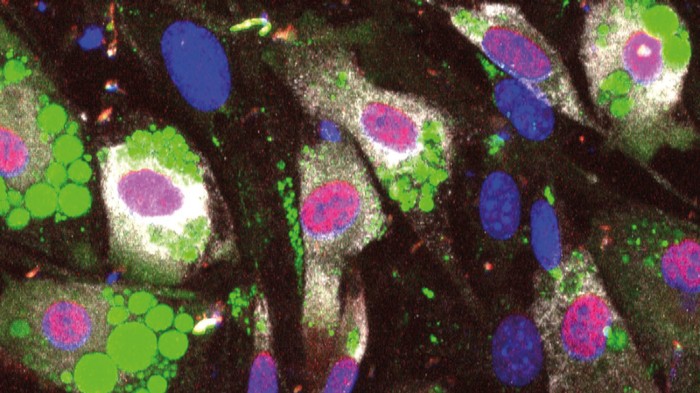
Single-cell immunofluorescence assay showing precursor cells maturing into adipose cells full of fat

Precursor cells (blue) mature into adipose cells full of fat (green) with help from hormones, whose levels surge and fall over a day. Credit: Z. Bahrami-Nejad et al/Cell Metab.

PHYSIOLOGY


Why fat piles on when the body's daily cycles are in disarray

Timing of hormone fluctuations influences fat cells' development.

Changes in the patterns of hormone production might cause weight gain when circadian rhythms are disrupted.

Hormones called glucocorticoids stimulate the production of mature fat cells. In humans, glucocorticoid levels naturally rise in the morning and fall in the evening, but stress can also elevate them.

To study how glucocorticoid levels relate to weight gain, Mary Teruel at Stanford University in California and her colleagues injected mice with glucocorticoids at varying times of day, but fed all mice the same amount of food. Mice given the hormone late in their wakeful phase gained weight. But mice injected just after they'd woken up — when their glucocorticoid levels were already naturally high — did not.

The results suggest that high glucocorticoid levels at unusual times of day could contribute to weight gain. This could help to explain why stress and disrupted sleep cycles are linked to rising weight.


Benjamin Hidalgo-Matlock
Skin Care Physicians of Costa Rica
4000-1054
2208-8206
Please excuse the shortness of this message, as it has been sent from a mobile device.

Thursday, April 12, 2018

Manchas en manos

https://www.epocrates.com/sites/default/files/res/dacc/2018/PellagraRashBMJ1803.pdf

Wednesday, April 11, 2018

Tratamiento con microagujas para vitiligo

Comparison between the efficacy of microneedling combined with 5-fluorouracil vs microneedling with tacrolimus in the treatment of vitiligo.

Mina M, et al. J Cosmet Dermatol. 2018.

Authors

Author information

1
Faculty of Medicine, Department of Dermatology & Venereology, Tanta University, Tanta, Egypt.
2
Faculty of Medicine, Department of pathology, Tanta University, Tanta, Egypt.

Citation

J Cosmet Dermatol. 2018 Mar 12. doi: 10.1111/jocd.12440. [Epub ahead of print]

Abstract

BACKGROUND: Several treatment modalities had been used for the treatment of vitiligo, but the optimal treatment has not yet been identified.

OBJECTIVES: To study the efficacy of microneedling with 5-flurouracil vs its efficacy with tacrolimus in the treatment of vitiligo.

PATIENTS AND METHODS: Twenty-five patients with vitiligo were subjected to microneedling of 2 patches of vitiligo with dermapen, then application of 5-fluorouracil to 1 patch and tacrolimus on the other patch. This procedure was repeated every 2 weeks for every patient for maximum 6 months (12 sessions). The patients were followed up for 3 months after the last session.

RESULTS: The overall repigmentation was significantly higher in 5-fluorouracil-treated patches compared with tacrolimus. Excellent improvement occurred in 48% of 5- flurouracil-treated patches while only in 16% of tacrolimus-treated patches. In the acral parts, 40% of the patches treated with 5-fluorouracil achieved excellent improvement (repigmentation >75%), while no patch in the acral parts achieved excellent improvement with tacrolimus. However, there was significant difference between the 2 drugs,regarding inflammation, ulceration, and hyperpigmentation which occurred with 5-fluorouracil.

CONCLUSION: Microneedling combined with 5-fluorouracil or tacrolimus is safe and effective treatment of vitiligo. However, 5-fluorouracil achieved a greater percentage of repigmentation than tacrolimus particularly in the acral parts.

© 2018 Wiley Periodicals, Inc.

PMID

 29532621 [ - as supplied by publisher]


Benjamin Hidalgo-Matlock
Skin Care Physicians of Costa Rica
4000-1054
2208-8206
Please excuse the shortness of this message, as it has been sent from a mobile device.

Tuesday, April 10, 2018

Varios de Clubdermaweb...

BIBLIOGRAPHIC SURVEY N°101

MICRONEEDLING IN THE TREATMENT OF PIGMENTED POST ACNE SCARRING IN DARKER SKIN

This study aimed to evaluate the efficacy and safety of the use of microneedling for the treatment of hyperpigmented acne scars in patients with darker skin types.

Thirty-nine patients with skin type III, IV and V (Fitzpatrick) were included. There was an initial evaluation of both acne scars and pigmentation indexes using PAHPI (post-acne hyperpigmentation index) and Goodman and Baron scale (GB scale). Treatment with microneedle was performed and patients were assessed after 2 and 4 weeks. Photos were taken at baseline and at the final evaluation for comparison.

Results indicated a statistically significant improvement in both the PAHPI index and Goodman and Baron scale. The adverse effects observed were transient erythema, mild dryness and small bruising and there was no pigmentation exacerbation in any of the patients.

The authors concluded that use of microneedling for the treatment of hyperpigmented acne scars in higher skin phototypes is effective and safe, presenting only mild and transient adverse effects. It was pointed out that some patients may require complementary treatments to improve hyperpigmentation.

J Cosmet Dermatol. 2018 Mar 15. doi: 10.1111/jocd.12520. [Epub ahead of print]
Skin microneedling for acne scars associated with pigmentation in patients with dark skin.
Al Qarqaz F, Al-Yousef A.

IDIOPATHIC GUTTATE HYPOMELANOSIS - TREATMENT WITH FRACTIONAL ABLATIVE PHOTOTHERMOLYSIS VERSUS TOPICAL RETINOID

This prospective study compared the efficacy of idiopathic guttate hypomelanosis treatment with fractional CO2 laser (CO2FL), Er: YAG fractional laser (EYFL) and topical use of 0.025% tretinoin.

A total of 122 lesions of 18 patients (mean age 70 ± 20 years) were analyzed. The lesions were distributed among 3 treatment groups: A. (n = 42) CO2FL, B. (n = 40) EYFL and C. (n = 40) topical tretinoin. Results were evaluated using Visual Analogue Scale (VAS) - measuring the severity of the lesion, Investigator's Global Assessment (IGA) - checking the extent of improvement achieved by the treatment, and Patient's Satisfaction Score (PSS).

The results indicated significantly superior efficacy with laser treatments (CO2FL and EYFL) compared to topical tretinoin treatment (p <0.01 in VAS, IGA and PSS). There was, however, no significant difference between the results obtained with both fractional lasers.

The authors concluded that fractional ablative lasers are effective in the treatment of idiopathic guttate hypomelanosis, showing results in a short period of time and without any serious adverse effects.

.

J Cosmet Laser Ther. 2018 Mar 16:1-5. doi: 10.1080/14764172.2018.1444771. [Epub ahead of print]
Comparative study of ablative fractional photothermolysis versus topical retinoid cream in the treatment of idiopathic guttate hypomelanosis.
Koh WS, Kim JE, Ro YS, Ko JY.

INTRADERMAL TRANEXAMIC ACID TO PREVENT POST-INFLAMMATORY HYPERPIGMENTATION AFTER SOLAR LENTIGINES REMOVAL

This study evaluated safety and efficacy of intradermal tranexamic acid (TA) injections after treatment of solar lentigines with Q-switched 532nm Nd: YAG laser, aiming at reducing the risks of post-inflammatory hyperpigmentation (PIH).

Twenty-five patients presenting 50 solar lentigines in the forearms were included. All selected lesions were submitted to QS 532nm Nd: YAG laser treatment. After the laser treatment, intradermal injections of TA 50mg / mL were applied in one lesion and saline solution (0.9%) in the other lesion. Assessment using Mexameter® was performed and two blinded dermatologists examined photos taken at baseline and after 2, 4, 8 and 12 weeks. Side effects observed with TA were minimal, with resolution within 1 hour.

Results at the end of the study indicated a significant reduction of the mean melanin index (MI) in both groups. At week 4 assessment, TA group presented a significant reduction in the mean MI compared to the control group (p = 0.025). Overall PIH rates were 16% for the TA group and 28% for the control group.

The authors concluded that TA (50m/mL) intradermal injections may reduce the risk of post-inflammatory hyperpigmentation 4 weeks after treating solar lentigines with QS 532nm Nd: YAG laser.

J Cosmet Laser Ther. 2018 Mar 5:1-7. doi: 10.1080/14764172.2018.1444770. [Epub ahead of print]
Intradermal tranexamic acid injections to prevent post-inflammatory hyperpigmentation after solar lentigo removal with a Q-switched 532-nm Nd:YAG laser.
Sirithanabadeekul P(1), Srieakpanit R(1).