Dermatología en Costa Rica

Monday, January 25, 2021

Prurito genitales y perianal

Journal Scan / Research · January 24, 2021

Iatrogenic ACD in the Perianal and Genital Area

Contact Dermatitis

TAKE-HOME MESSAGE


Abstract

Contact Dermatitis
Iatrogenic Allergic Contact Dermatitis in the (Peri)Anal and Genital Area
Contact Derm 2020 Dec 22;[EPub Ahead of Print], L Gilissen, I Schollaert, S Huygens, A Goossens

Sunday, January 24, 2021

Ocular Comorbidities in Patients With Hidradenitis Suppurativa


TAKE-HOME MESSAGE


Abstract 


Hidradenitis suppurativa (HS) is a chronic systemic inflammatory disorder of the terminal follicular epithelium in the apocrine gland–bearing skin. Clinically, it is characterized by lesions, including comedones, papules, pustules, nodules, and fistulas, that are mainly concentrated in axillae, perineal, and submammary areas.1

Other autoimmune diseases are associated with ocular comorbidities; however, there is a paucity of data on ocular comorbidities in HS.1-4 In this article, we aim to quantify the burden of ocular comorbidities in patients with HS.


JAMA Dermatology

Burden of Ocular Comorbidities in Patients With Hidradenitis Suppurativa

JAMA Dermatol 2021 Jan 06;[EPub Ahead of Print], RRZ Conic, G Fabbrocini, C Marasca, NL Bragazzi, W Abdulla, M Adawi, G Damiani 


Sent from my iPhone

Benjamin Hidalgo-Matlock
Skin Care Physicians of Costa Rica

Clinica Victoria en San Pedro: 4000-1054
Momentum Escazu: 2101-9574

Please excuse the shortness of this message, as it has been sent from
a mobile device.

Friday, January 22, 2021

Risk Factors for Surgical Site Infection After Minor Dermatologic Surgery Advances in skin & wound care

TAKE-HOME MESSAGE

Abstract


BACKGROUND

Surgical site infection (SSI) after dermatologic surgery is associated with poor outcomes including increased recovery time, poor cosmesis, and repeat visits to doctors. Prophylactic antibiotics are prescribed to reduce these adverse outcomes. Identifying risk factors for SSI will facilitate judicious antibiotic prophylaxis.

OBJECTIVE

To identify risk factors for SSI after minor dermatologic surgery.

METHODS

Individual patient data from four large randomized controlled trials were combined to increase statistical power. A total of 3,819 adult patients requiring minor skin procedures at a single facility were recruited over a 10-year period. The main outcome measure was SSI.

MAIN RESULTS

A total of 298 infections occurred, resulting in an overall incidence of 7.8% (95% confidence interval [CI], 5.8–9.6), although the incidence varied across the four studies (P = .042). Significant risk factors identified were age (relative risk [RR], 1.01; 95% CI, 1.001–1.020; P = .008), excisions from the upper limbs (RR, 3.03; 95% CI, 1.76–5.22; P = .007) or lower limbs (RR, 3.99; 95% CI, 1.93–8.23; P = .009), and flap/two-layer procedures (RR, 3.23; 95% CI, 1.79–5.85; P = .008). Histology of the excised lesion was not a significant independent risk factor for infection.

CONCLUSIONS

This study demonstrated that patients who were older, underwent complex excisions, or had excisions on an extremity were at higher risk of developing an SSI. An awareness of such risk factors will guide evidence-based and targeted antibiotic prophylaxis.


Advances in skin & wound care
Risk Factors for Surgical Site Infection After Minor Dermatologic Surgery
Adv Skin Wound Care 2021 Jan 01;34(1)43-48, M Delpachitra, C Heal, J Banks, D Charles, S Sriharan, P Buttner 


Sent from my iPhone

Benjamin Hidalgo-Matlock
Skin Care Physicians of Costa Rica

Clinica Victoria en San Pedro: 4000-1054
Momentum Escazu: 2101-9574

Please excuse the shortness of this message, as it has been sent from
a mobile device.

Thursday, January 14, 2021

Journal Scan / Case Study · January 15, 2021 Occipital Fibrosing Alopecia International Journal of Dermatology


TAKE-HOME MESSAGE


Abstract 

Frontal fibrosing alopecia (FFA) classically results in a band‐like scarring alopecia involving the frontotemporal hairline and eyebrows.1-4 Diagnosis is confirmed by trichoscopy showing the absence of vellus hairs and peripilar casts and pathology showing scarring alopecia with a lichenoid infiltrate. Occipital involvement has been described in up to 32% of cases; however, it usually occurs in association with frontoparietal scarring.5 We report here a case series of 14 women with FFA limited to the occipital scalp.

International Journal of Dermatology
Occipital Fibrosing Alopecia
Int. J. Dermatol 2020 Dec 20;[EPub Ahead of Print], MA Martinez-Velasco, B Burroway, C Thompson, T Silyuk, A Tosti 

Sent from my iPhone

Benjamin Hidalgo-Matlock
Skin Care Physicians of Costa Rica

Clinica Victoria en San Pedro: 4000-1054
Momentum Escazu: 2101-9574

Please excuse the shortness of this message, as it has been sent from
a mobile device.

Monday, January 11, 2021

Covid-19 y panza

Gut Microbiome Altered in COVID-19 Patients

By Kelly Young

Edited by David G. Fairchild, MD, MPH, and Jaye Elizabeth Hefner, MD

The gut microbiome appears to be altered in patients with COVID-19, finds a study in Gut.

Researchers compared blood and stool samples of roughly 100 patients with COVID-19 in Hong Kong with samples from other people taken before the pandemic. Nearly half the COVID-19 patients had mild disease.

Several bacteria species were associated with disease severity after adjustment for antibiotic use. For instance, compared with controls, patients with COVID-19 had smaller populations of Faecalibacterium prausnitzii and Eubacterium rectale, which have known immunomodulatory potential. Even among those who'd recovered from COVID-19, the gut remained distinct from non-COVID patients a median of 6 days after testing negative.

The authors say that their results "suggest that gut microbiota composition is associated with the magnitude of immune response to COVID-19 and subsequent tissue damage and thus could play a role in regulating disease severity." They add, "Bolstering of beneficial gut species depleted in COVID-19 could serve as a novel avenue to mitigate severe disease, underscoring importance of managing patients' gut microbiota during and after COVID-19."

Gut article (Free abstract)

NEJM Journal Watch COVID-19 page (Free)

NEJM COVID-19 page (Free)

Wednesday, January 06, 2021

PCR para escabiosis

TCHING TO SEE IF POLYMERASE CHAIN REACTION MITE BE THE FUTURE OF DIAGNOSING SCABIES


DII small banner

By Warren R. Heymann, MD
January 6, 2021
Vol. 3, No. 1

The scourge of scabies has been plaguing mankind for millennia. The causal relationship of the itch mite causing scabies was discovered by Bonomo and Cestoni in 1687, earning the distinction of being the first to discover a proven etiology for a human disease. (1)

Scabies is an infestation of the skin by the miteSarcoptes scabiei. Transmission is by direct skin-to-skin contact or indirectly through fomites. Symptoms typically appear 3 to 6 weeks after an infestation. In patients with a prior exposure to the mite, symptoms can appear as early as 24 hours post exposure. Lesions consist of pruritic erythematous papules with excoriations. They're usually symmetrical and involve the interdigital webs, the flexural aspect of wrists, the axillae, the peri-umbilical area, elbows, buttocks, feet, genital area in men, and the peri-areolar area in women (called the circle of Hebra). The entire integument, including the face and the scalp, can be involved in infants, the elderly, and immunosuppressed patients. The pathognomonic sign is the burrow, representing the tunnel that the female mite digs to lay her eggs. Crusted scabies (CS) is a severe form that occurs in immunosuppressed individuals such as patients with AIDS, manifesting with extensive hyperkeratosis, predominantly over the scalp and the extremities. (2)

Illustration for DWII PCR scabies
JAAD 2008; 59: 530.

The brilliance of Bonomo and Cestoni is magnified by my own difficulty in identifying mites, eggs, or scybala within skin scrapings. Thinking of scabies in a differential diagnosis is easy (except in atypical cases!) — securing the diagnosis is another issue. 

A Delphi study of the International Alliance for the Control of Scabies (IACS) has proposed the following criteria for the diagnosis of scabies (the diagnosis may be rendered on any of the levels; for levels B and C, scabies should be diagnosed after ruling out other disorders in the differential diagnosis) (3):

  • A: Confirmed scabies having at least one of: (A1) — mites, eggs, or feces on light microscopy of skin samples; (A2) — mites, eggs, or feces visualized on individual using a high-powered imaging device; (A3) — mite visualized on individual using dermoscopy.

  • B: Clinical scabies having at least one of: (B1) — scabies burrows; (B2) — typical lesions affecting male genitalia; (B3) — typical lesions in a typical distribution and two history features (itch or close contact with pruritus or a typical case of scabies).

  • C: Suspected scabies having one of: (C1) — typical lesions in a typical distribution and one history feature; (C2) — atypical lesions or atypical distribution and two history features.

I am almost embarrassed to admit it, but I would add a "D" category — pruritic patients with no obvious lesions or laboratory findings, for whom I prescribe either empiric permethrin or oral ivermectin, hoping that it would cure the patient. I'd be willing to wager that I'm not the only dermatologist who has resorted to this dermatological chicanery. 

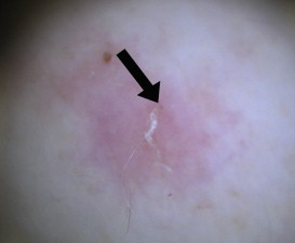
Using mineral oil scrapings may be the gold standard for diagnosis, but it takes skill. One of my partners is particularly adept at finding mites, while I often struggle with these preparations. I feel more confident with dermoscopy, looking for the "delta" (the dark brown triangular structure corresponding to the mite's anterior at the end of the burrow). Others advocate the burrow ink test. (4,5)

In that context, I was most intrigued by the study by Delaunay et al who evaluated polymerase chain reaction (PCR) as a diagnostic method for scabies using nonexpert-dependent standardized sampling.

Dry swabs were systematically rubbed across the front of both wrists, the eight interdigital spaces, and on any suspected scabies lesions in all patients referred for scabies. A new PCR-based diagnostic test was run on the samples. All patients underwent clinical and dermoscopic examination. Scabies diagnosis was confirmed when dermoscopic examination was positive or the patient had typical clinical signs of scabies. A total of 164 patients were sampled; 87 had confirmed scabies and 77 did not. Of the 87 patients with proved scabies, 33 patients had positive scabies PCR, resulting in a 37.9% sensitivity and a 61.7% negative predictive value. None of the 77 patients ruled out for scabies had a positive PCR result. The authors concluded that scabies PCR is specific but has poor sensitivity. Regardless, this PCR diagnostic method based on nontraumatic standardized skin sampling is not expert-dependent and is reproducible. This diagnostic method may be relevant as a non-expert sentinel diagnosis tool in large clusters where a scabies outbreak is suspected, such as nursing homes or other long-term care facilities. (6)

As much as I enjoy my consultative practice, I would be fine without seeing patients sent from facilities with the only clinical information being "rule out scabies." I'm itching for the development of a PCR analysis with a higher sensitivity for scabies — that would be a boon for institutional administrators, practitioners, and patients alike. 

Point to Remember: Scabies may be the easiest or hardest diagnosis to render. Perhaps development of novel techniques such as PCR will simplify the process.

Our Expert's Viewpoint

Aileen Y. Chang, MD
Assistant Clinical Professor of Dermatology
University of California, San Francisco 
International Alliance for the Control of Scabies (
www.controlscabies.org)

In this day and age, it is hard to believe that we still do not have a sensitive point-of-care test for scabies. While the diagnostic PCR method published by Delauney et al. will not help us with diagnosing individual patients with scabies, it may be a potential diagnostic method for settings where the diagnosis of one individual with scabies would result in treatment of many other individuals. These settings include long-term care facilities, prisons, and childcare facilities, among others. Most of these facilities do not have quick access to a scabies expert and when faced with a potential scabies outbreak, they need to make an informed decision on whether or not to treat. In a large facility, treatment for scabies is expensive and time-intensive, requires development of a structured implementation plan, and can have a significant toll on staff and facility residents. The decision to treat scabies in these settings cannot be taken lightly. This diagnostic method has a sensitivity of 90.8% with five samples and 99.1% with 10 samples. As the authors conclude, their findings need to be validated in the context of an outbreak and with adults, as most individuals in the study were children. Delauney et al.'s work is a step in the right direction. 

  1. Arlian LG, Morgan MS. A review of Sarcoptes scabiei: Past, present, and future. Parasit Vectors 2017; 10: 297. 

  2. Khalil S, Abbas O, Kibbi AG, Kurban M. Scabies in the age of increasing drug resistance. PLoS Negl Trop Dis 2017: 11 (11): e0005920

  3. Engelman D. Fuller LC, Steer AC, International Alliance for the Control of Scabies Delphi panel. Consensus criteria for the diagnosis of scabies: A Delphi study of international experts. PLoS Negl Trop Dis 2018;12(5):e0006549

  4. Tang J, You Z, Ran Y. Simple methods to enhance the diagnosis of scabies. J Am Acad Dermatol 2019; 80: e99-100.

  5. Leung V, Miller M. Detection of scabies: A systematic review of diagnostic methods. Can J Infect Dis Med Microbiol 2011; 22: 143-146. 

  6. Delaunay P, Hérissé AL, Hasseine L, Chiaverini C, et al. Scabies polymerase chain reaction with standard dry swab sampling: An easy tool for cluster diagnosis of human scabies. Br J Dermatol 2020; 182: 197-201. 


All content found on Dermatology World Insights and Inquiries, including: text, images, video, audio, or other formats, were created for informational purposes only. The content represents the opinions of the authors and should not be interpreted as the official AAD position on any topic addressed. It is not intended to be a substitute for professional medical advice, diagnosis, or treatment.

DW Insights and Inquiries archive

Explore hundreds of Dermatology World Insights and Inquiries articles by clinical area, specific condition, or medical journal source


Sent from my iPhone

Benjamin Hidalgo-Matlock
Skin Care Physicians of Costa Rica

Clinica Victoria en San Pedro: 4000-1054
Momentum Escazu: 2101-9574

Please excuse the shortness of this message, as it has been sent from
a mobile device.

Tuesday, January 05, 2021

Member-Membership-Benefits-Fellow-logo-Logos-AAD-Fellow-logo-black-HiRes.jpg 1.842×417 pixels

Saturday, January 02, 2021

Use of Hydroxychloroquine and Risk of Major Adverse Cardiovascular Events in Patients With Lupus Erythematosus


TAKE-HOME MESSAGE


Abstract


BACKGROUND

Limited data suggest that hydroxychloroquine may affect risk of cardiovascular disease in patients with lupus erythematosus (LE).

OBJECTIVE

To investigate whether hydroxychloroquine treatment is associated with major adverse cardiovascular events (MACE) (myocardial infarction, ischemic stroke, or cardiovascular-associated death) in patients with cutaneous LE (CLE) or systemic LE (SLE).

METHODS

Based on the Danish nationwide registers, an observational cohort study was conducted including patients with first-time diagnosis of CLE or SLE (between 1997-2017). Cox regression models calculating hazard ratio (HR) analyzing the risk of MACE was performed comparing time on and off hydroxychloroquine (including never-user). The models were adjusted for age, sex, socio-economic status, concomitant treatment, and cardiovascular risk factors.

RESULTS

Among 4587 patients with LE, 51% (n=2343) were treated with HCQ during the study period. An inverse association between use of HCQ and MACE risk was observed among patients with SLE (adjusted HR 0.65; 95% CI 0.46-0.90) and patients with CLE (adjusted HR 0.71; 95% CI 0.42-1.19). Consistent results were found in sensitivity analyses including a case-time-control design.

LIMITATIONS

No information on disease activity/severity was available.

CONCLUSION

Our findings indicate an opportunity to reduce risk of cardiovascular events in patients with LE through use of hydroxychloroquine.


Journal of the American Academy of Dermatology
Use of Hydroxychloroquine and Risk of Major Adverse Cardiovascular Events in Patients With Lupus Erythematosus: A Danish Nationwide Cohort Study
J Am Acad Dermatol 2020 Dec 12;[EPub Ahead of Print], JH Haugaard, L Dreyer, MB Ottosen, G Gislason, K Kofoed, A Egeberg


Sent from my iPhone

Benjamin Hidalgo-Matlock
Skin Care Physicians of Costa Rica

Clinica Victoria en San Pedro: 4000-1054
Momentum Escazu: 2101-9574

Please excuse the shortness of this message, as it has been sent from
a mobile device.

Vaccination Against HPV JAMA Dermatology

TAKE-HOME MESSAGE


Abstract 


CLINICAL QUESTION

Who should receive human papillomavirus (HPV) vaccination?

BOTTOM LINE

Catch-up vaccination is now recommended for all persons through age 26 years. For persons aged 27 through 45 years, clinicians and patients should now jointly decide whether HPV vaccination is appropriate. Routine HPV vaccination at age 11 or 12 years (or as early as age 9 years) continues to be recommended.


JAMA Dermatology
Vaccination Against Human Papillomavirus
JAMA Dermatol 2020 Aug 19;[EPub Ahead of Print], DH Marks, KA Katz 


Sent from my iPhone

Benjamin Hidalgo-Matlock
Skin Care Physicians of Costa Rica

Clinica Victoria en San Pedro: 4000-1054
Momentum Escazu: 2101-9574

Please excuse the shortness of this message, as it has been sent from
a mobile device.

Staphylococcal Scalded Skin Syndrome Pediatric Dermatology

TAKE-HOME MESSAGE


Abstract


BACKGROUND

Staphylococcal scalded skin syndrome (SSSS) is a toxin-mediated, blistering skin disorder that mainly affects infants and children. There is limited literature regarding pediatric SSSS. The purpose of this study was to describe the epidemiology, clinical features, and management of pediatric SSSS.

METHODS

Retrospective cohort study of pediatric patients with a clinical diagnosis of SSSS seen at the Hospital for Sick Children in Toronto, Ontario, Canada, from January 1994 to March 2016.

RESULTS

We included 84 patients with a clinical diagnosis of SSSS; 49/84 (58%) were male. Mean age of diagnosis was 3.1 ± 2.4 years. All patients presented with erythema and exfoliation, while 64/84 (76%) presented with vesicles/ bullae. Skin tenderness was the most common symptom, present in 68/84 (81%) subjects. Staphylococcus aureus was more commonly isolated from periorificial cultures than from bullae. Mean hospitalization was 4.7 ± 2.3 days. No difference was found in admission duration between children receiving clindamycin and those that did not (3.6 ± 2.2 vs 3.9 ± 2.34 days, P = .63). Skin debridement was the only risk factor leading to more complications and prolonged hospitalization (P = .03). Severe complications were seen in 4 (5%) cases, and no fatalities were observed.

CONCLUSIONS

Healthcare providers should be aware of SSSS and consider it in the differential diagnosis of infants and children with new onset erythema, exfoliation, and/or vesiculation. Suspected culprit pathogens were more often obtained from periorificial swabs; however, these isolates were not tested for exfoliative toxin to confirm causality. Antibiotic treatment should be guided by sensitivity testing. Addition of clindamycin as an anti-toxin agent had no effect on the duration of hospitalization, and this should be further investigated. Surgical debridement of the skin in patients with SSSS should be discouraged.


Pediatric Dermatology
Staphylococcal Scalded Skin Syndrome: An Epidemiological and Clinical Review of 84 Cases
Pediatr Dermatol 2020 Dec 01;[EPub Ahead of Print], C Liy-Wong, E Pope, M Weinstein, I Lara-Corrales 

Sent from my iPhone

Benjamin Hidalgo-Matlock
Skin Care Physicians of Costa Rica

Clinica Victoria en San Pedro: 4000-1054
Momentum Escazu: 2101-9574

Please excuse the shortness of this message, as it has been sent from
a mobile device.