Dermatología en Costa Rica

Saturday, February 27, 2021

Topical Timolol 0.5% Gel-Forming Solution for Erythema in Rosacea


TAKE-HOME MESSAGE


Abstract 

Current topical medications for rosacea-associated erythema include brimonidine and oxymetazoline, which induce cutaneous vasoconstriction. Topical timolol has been used for infantile hemangioma due to its antiangiogenic and vasoconstrictive effects, but its efficacy for treating rosacea-associated erythema is unknown

Journal of the American Academy of Dermatology
Topical Timolol 0.5% Gel-Forming Solution for Erythema in Rosacea: A Quantitative, Split-Face, Randomized, and Rater-Masked Pilot Clinical Trial
J Am Acad Dermatol 2021 Feb 03;[EPub Ahead of Print], J Tsai, AL Chien, N Kim, S Rachidi, BM Connolly, H Lim, SS Alessi César, S Kang, LA Garza 


Sent from my iPhone

Benjamin Hidalgo-Matlock
Skin Care Physicians of Costa Rica

Clinica Victoria en San Pedro: 4000-1054
Momentum Escazu: 2101-9574

Please excuse the shortness of this message, as it has been sent from
a mobile device.

Friday, February 26, 2021

Antigeno Candida para Verrugas Periungeales

Journal Scan / Research · February 24, 2021

Intralesional Antigen Immunotherapy for Treatment of Periungual Warts

TAKE-HOME MESSAGE

Abstract


BACKGROUND

Intralesional immunotherapy using different types of antigens is considered an effective and safe treatment option for different types of warts. However, there are few studies that illustrate the use of these antigens in the treatment of periungual warts as a distinct type of warts.

OBJECTIVE

To evaluate the efficacy and safety of three antigens: measles, mumps, rubella (MMR) vaccine, Candida antigen, and purified protein derivative (PPD) in the treatment of periungual warts.

METHODS

The study included 150 patients who were randomly assigned to 3 groups with 50 patients in each. Each agent was injected intralesionally at a dose of 0.1 mL into the largest wart at 2-week intervals until complete clearance or for a maximum of 5 sessions.

RESULTS

Complete clearance of warts was observed in 70%, 80%, and 74% in PPD, Candida antigen, and MMR vaccine groups, respectively. There was no statistically significant difference regarding the therapeutic response between the 3 studied groups. Adverse effects were transient and insignificant in the 3 groups. No recurrence of the lesions was reported in any of the studied groups.

CONCLUSIONS

Intralesional antigen immunotherapy seems to be an effective therapeutic option for the treatment of periungual warts.


Journal of cutaneous medicine and surgery
Intralesional Antigen Immunotherapy in the Treatment of Periungual Warts
J Cutan Med Surg 2021 Jan 27;[EPub Ahead of Print], A Nofal, R Alakad, I Fouda, MM Fawzy

Sent from my iPhone

Benjamin Hidalgo-Matlock
Skin Care Physicians of Costa Rica

Clinica Victoria en San Pedro: 4000-1054
Momentum Escazu: 2101-9574

Please excuse the shortness of this message, as it has been sent from
a mobile device.

Wednesday, February 24, 2021

Effect of Immunosuppression on Patch Testing in Patients With IBD


TAKE-HOME MESSAGE

The Effect of Immunosuppression on Patch Testing: A Cross-Sectional Study in Patients With Inflammatory Bowel Disease
Contact Derm 2021 Jan 13;[EPub Ahead of Print], A Flynn, M Malik, S McCarthy, F Shanahan, J Bourke 


Sent from my iPhone

Benjamin Hidalgo-Matlock
Skin Care Physicians of Costa Rica

Clinica Victoria en San Pedro: 4000-1054
Momentum Escazu: 2101-9574

Please excuse the shortness of this message, as it has been sent from
a mobile device.


Sent from my iPhone

Benjamin Hidalgo-Matlock
Skin Care Physicians of Costa Rica

Clinica Victoria en San Pedro: 4000-1054
Momentum Escazu: 2101-9574

Please excuse the shortness of this message, as it has been sent from
a mobile device.

Sunday, February 21, 2021

Tx escabiasis menores de 2 meses

Management of Scabies in Infants Younger Than 2 Months

TAKE-HOME MESSAGE


Abstract 

BACKGROUND/OBJECTIVES

Efficacy and safety data of scabies treatments in infants are limited. Although topical permethrin is used in the treatment of scabies in adults, it is not approved for use in infants younger than 2 months of age in many parts of the world. This study aimed to describe treatment practices in the management of scabies in infants younger than 2 months.

METHODS

An online survey was developed and distributed to physicians worldwide through the Society of Pediatric Dermatology and the Pediatric Dermatology Research Alliance. Data collected included demographics, medication availability, experience using medications, deterrents to medication use, medication administration preferences, perceived and experienced medication side effects, and preferred treatment agent in this population.

RESULTS

In total, 57 physicians from seven countries responded. The majority of respondents were board-certified in pediatric dermatology (48/57, 84.2%) and resided in the United States (44/57, 77.2%). Respondents had experience using permethrin (47/57, 82.5%) and precipitated sulfur (35/57, 61.4%) most frequently. Most (38/57, 66.7%) preferred permethrin as their treatment of choice. Among those who did not use permethrin, potential side effects (8/10, 80%) were most frequently reported as a deterrent from its use. However, only 4.3% (22/47) of those who used permethrin reported side effects, including itching, erythema, and xerosis.

CONCLUSIONS

Permethrin is frequently used in the treatment of infants younger than 2 months with scabies. Furthermore, our results demonstrate that permethrin is the preferred treatment agent among sampled dermatologists for infants younger than 2 months. Few side effects were reported, and none were serious.


Pediatric Dermatology
Treatment Practices in the Management of Scabies in Infants Younger Than Two Months
Pediatr Dermatol 2021 Jan 24;[EPub Ahead of Print], C Thomas, W Rehmus, AY Chang 


Sent from my iPhone

Benjamin Hidalgo-Matlock
Skin Care Physicians of Costa Rica

Clinica Victoria en San Pedro: 4000-1054
Momentum Escazu: 2101-9574

Please excuse the shortness of this message, as it has been sent from
a mobile device.

Friday, February 19, 2021

Angiomatosis dermica difusa... angiomatosis reactiva tuve un caso recientemente biopsia Granuloma Piogeno

Honing in on diffuse dermal angiomatosis


DII small banner

By Warren R. Heymann, MD
February 17, 2021
Vol. 3, No. 7

 Diffuse dermal angiomatosis (DDA) was conceptualized as a variant of reactive angioendothelimatosis (RA) in 1994 by Krell et al, reporting on two women, both with severe peripheral vascular disease requiring bypass grafts, presenting with ulcerated, violaceous plaques on the legs. The authors observed: "Unlike previously described cases of RA, our patients' lesions were due to a diffuse proliferation of endothelial cells in the reticular dermis with only minimal, focal proliferation of these cells. Positive immunostaining with antibodies to Factor VIII-related and CD34 antigens adds evidence that the proliferated cells in the dermis were endothelial cells." Both patients demonstrated complete clinical clearing after bypass grafts were placed suggesting that the inciting factor was vascular insufficiency. (1)

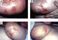
Aside from atherosclerosis, subsequent reports have demonstrated that DDA is associated with arteriovenous fistulae and patients with large, pendulous breasts (macromastia). My first introduction to DDA was a woman with ulcerations of her breasts — I recall that my clinical concern was either vasculitis or breast cancer. Hui et al have reported that DDA may mimic inflammatory breast carcinoma both clinically and on mammography. (2) Other entities entering the differential diagnosis of DDA include Kaposi sarcoma, acroangiodermatitis (pseudo-Kaposi sarcoma), angiosarcoma, (3) and any occlusive vasculopathy/vasculitis causing a retiform purpura. (4)

Other ischemic factors associated with DDA include smoking, cutis marmorata telangiectatica congenita, granulomatosis with polyangiitis, anticardiolipin antibodies, and calciphylaxis. (3) In their review of 24 cases of calciphylaxis, O'Connor et al identified seven cases of DDA. The only statistically significant associations in these cases were African-American race and congestive heart failure. (5)

Image for DWII on diffuse dermal angiomatosis Image from reference 7.

Grossman-Kranseler et al provide a cogent conceptualization of the pathophysiology of DDA. The common denominator in the pathophysiology of DDA is ischemia causing hypoxia leading to increased VEGF (vascular endothelial growth factor) yielding increased vascular proliferation. "Hypoxia inducible factor (HIF-1) stabilization is critical to wound healing and angiogenesis following hind limb ischemia models. HIF-1 stimulates the stromal derived factor-1 (SDF-1)/CXCR-4 axis, which stimulates the proliferation and homing of CD34+endothelial progenitor cells. VEGF contributes to endothelial cell differentiation. Likely, the CD34+/VEGF+ spindle cell population represents an endothelial progenitor cell population. Further histologic/morphologic and immunohistochemical studies could be performed in cases of DDA to determine the cell-specific expression of SDF-1, CXCR-4, and HIF-1." (6)

Treatment of DDA revolves around improving blood flow by judicious consideration of any of the following: cessation of smoking, revascularization (bypass grafts, stents), and anticoagulation. Steroids and isotretinoin have been reported to be effective based on their ability to inhibit angiogenesis. McLaughlin et al reported the case of a 28-year-old woman with ulcerative breast DDA, who improved dramatically on isotretinoin (1 mg/kg for 2 months) with complete healing of the ulcers. (7)

There is much to be learned about the molecular aspects of hypoxia, wounds, and wound healing. VEGF elevation has also been observed in other entities such as von Hippel-Lindau disease and POEMS syndrome. Bevacizumab infusions have safely extended survival in a report of two patients with otherwise untreatable, life-threatening POEMS syndrome. (8) To the best of my knowledge, bevacizumab has not been utilized in patients with DDA. I suspect it would be valuable in recalcitrant cases unresponsive to the previously mentioned therapeutic options.

Point to Remember: Diffuse dermal angiomatosis reflects elevated VEGF in response to hypoxia. It should be in the differential diagnosis of ulcerated plaques in the context of retiform purpura, especially in those with vascular compromise. The prognosis is good if reconstitution of vascular perfusion can be accomplished — by surgical or medical means.

Our expert's viewpoint

Jack L. Arbiser, MD, PhD
Thomas J. Lawley Professor of Dermatology
Emory University School of Medicine

Grossman-Kranseler et al describe a case of diffuse dermal angiomatosis (DDA), a rare vascular proliferative disorder in an unusual location. Notably, they exclude infectious etiologies (HHV8) and demonstrate the presence of the angiogenic factor vascular endothelial growth factor (VEGF) in the lesions. They hypothesize that hypoxia induces hypoxia inducible factor 1 (HIF1) leading to vascular proliferation. We previously described a case of ulcerated DDA which occurred in pendulous breast tissue that responded to isotretinoin, which has antiangiogenic activities.

While DDA is a rare disease, it sheds light on more common disorders, characterized by endothelial proliferation and ulceration. While ulceration is instinctively thought to be a disorder of insufficient vascularization, it is usually characterized by excess endothelial proliferation. It would be more accurate to call most ulceration a disorder of inefficient vascularization. The endothelial proliferation and inflammation results in an environment hostile to re-epithelialization. DDA is usually associated with defects in large vessel perfusion (pendulous breasts, fistulas, etc.) but ulceration can be associated with small vessel perfusion defects (hemangioma of infancy, pyogenic granuloma, venous ulcers). More recently, we have identified novel therapeutic strategies to address ulcerative disorders. These include NADPH oxidase inhibitors such as gentian violet, which has been used on hemangiomas of infancy and most recently in a series of patients with pyogenic granuloma. We have identified another growth factor associated with excess vascular proliferation, Angiopoeitin like 4 (Angptl4), that is a major growth factor in hemangiomas, and that it can be blocked by R-propranolol, an isomer of propranolol that is present in commercial propranolol, but is not a beta blocker. It is likely that R-propranolol is the active principle of propranolol in hemangiomas, and that Angptl4 is likely a major pathogenic factor in vascular proliferative diseases of the skin, including DDA.

  1. Krell JM, Sanchez RL, Solomon AR. Diffuse dermal angiomatosis : A variant of reactive cutaneous angioendotheliomatosis. J Cutan Pathol 1994; 21: 363-370.

  2. Hui Y, Elco CP, Heini NF, Lourenco AP, et al. Diffuse dermal angiomatosis mimicking inflammatory breast carcinoma. Breast j 2018; 24: 196-198.

  3. Touloei K, Tongdee E, Smirnov B, Nousari C. Diffuse dermal angiomatosis. Cutis 2019 ; 103 : 181-183.

  4. Bhattacharya T, Sluzevich J. Generalized retiform purpura as a presenting sign of diffuse dermal angiomatosis. Dermatol Online J 2018; 24 (5): 10.

  5. O'Connor HM, Wu Q, Lauzon SD, Forcucci JA. Diffuse dermal angiomatosis associated with calciphylaxis: A 5-year retrospective institutional review. J Cutan Pathol 2020; 47: 27-30.

  6. Grossman-Kranseler J, Rhim E, Ruhoy S. Diffuse dermal angiomatosis: Report of a classic case with a comment on the pathophysiology based on the histologic findings. Am J Dermatopathol 2019 Dec 11 [Epub ahead of print].

  7. McLaughlin ER, Morris R. Weiss SW, Arbiser JL. Diffuse dermal angiomatosis of the breast: Response to isotretinoin. J Am Acad Dermatol 2001; 45: 462-465.

  8. Perfetti V, Palladini G, De Amici M, Livraghi L, et al. Bevacizumab treatment followed by maintenance in life-threatening POEMS syndrome. Ann Hematol 2013; 92: 1133-1134.

Tuesday, February 16, 2021

Terbinafina y labs

Terbinafine for onychomycosis

Dr. Kirby also discussed the rationale for ordering regular LFTs in patients taking terbinafine for onychomycosis. "There is a risk of drug-induced liver injury from taking terbinafine, but it's rare," she said. "Can we be thoughtful about which patients we expose?"

Evidence suggests that patients with hyperkeratosis greater than 2 mm, with nail matrix involvement, with 50% or more of the nail involved, or having concomitant peripheral vascular disease and diabetes are recalcitrant to treatment with terbinafine

(J Am Acad Dermatol. 2019 Apr;80[4]:853-67). "If we can frame this risk, then we can frame it for our patients," she said. "We're more likely to cause liver injury with an antibiotic. When it comes to an oral antifungal, itraconazole is more likely than terbinafine to cause liver injury. The rate of liver injury with terbinafine is only about 2 out of 100,000. It's five times more likely with itraconazole and 21 times more likely with Augmentin."

She recommends obtaining a baseline LFT in patients starting terbinafine therapy "to make sure their liver is normal from the start." In addition, she advised, "let them know that there is a TB seroconversion risk of about 1 in 50,000 people, and that if it happens there would be symptomatic changes. They would maybe notice pruritus and have a darkening in their urine, and they'd have some flu-like symptoms, which would mean stop the drug and get some care."

Dr. Kirby emphasized that a patient's propensity for developing drug-induced liver injury from terbinafine use is not predictable from LFT monitoring. "What you're more likely to find is an asymptomatic LFT rise in about 1% of people," she said.

She disclosed that she has received honoraria from AbbVie, ChemoCentryx, Incyte, Janssen, Novartis, and UCB Pharma.


Sent from my iPhone

Benjamin Hidalgo-Matlock
Skin Care Physicians of Costa Rica

Clinica Victoria en San Pedro: 4000-1054
Momentum Escazu: 2101-9574

Please excuse the shortness of this message, as it has been sent from
a mobile device.

Risk of Systemic Infections Requiring Hospitalization in Children With AD

TAKE-HOME MESSAGE


Abstract 


BACKGROUND

Certain infections can trigger worsening of atopic dermatitis (AD). Further, an increased risk of extra-cutaneous infections has been suggested among children with AD, but data are sparse.

OBJECTIVES

To examine whether hospital managed pediatric AD is associated with increased risk of extra-cutaneous infections in childhood requiring hospitalisation.

METHODS

Nationwide based cohort study using Danish registries. All children aged under 18 years with a hospital in- or outpatient diagnosis of AD and reference children without hospital diagnosis of AD were matched on gender and age at first AD diagnosis date (1:10). Children were included from January 1, 1995 to December 31, 2016 and followed up until December 31, 2017. Study outcomes were extra-cutaneous infections that lead to hospitalisation. The severity of AD was defined by dispensations of AD treatments.

RESULTS

Of 19,415 AD children (median follow-up 7.4, interquartile range (IQR) 3.3-13.3 years) and 194,150 non-AD children (median follow-up 7.7, IQR 3.6-13.5 years), 56% were boys and 50% aged under 2 years. In adjusted analyses, children with AD had increased occurence of lower respiratory (aHR (adjusted hazard ratio)=1.79, 95% confidence interval (CI95%) 1.65-1.94), upper respiratory (aHR=1.59, CI95% 1.34-1.88), urinary tract (aHR=1.34 CI95% 1.16-1.54), musculoskeletal (aHR=1.33 CI95% CI95% 1.06-1.66), and gastrointestinal infections (aHR=1.24, CI95% 1.14-1.35), compared with children without a hospital diagnosis of AD. The associations did not clearly vary with the AD severity. The absolute risk difference per 10,000 person-years was 26.4 (CI95% 23.0-29.8) for lower respiratory, 3.1 (CI95% 1.6-4.7) for upper respiratory, 3.6 (CI95% 1.8-5.4) for urinary tract, 0.9 (CI95% 0.2-2.0) for musculoskeletal and 8.7 (CI95% 5.7-11.7) for gastrointestinal infections.

CONCLUSIONS

Children with hospital-managed AD have an increased risk of systemic infections that lead to hospitalization. The absolute risk was generally low.


The British Journal of Dermatology
The Risk of Systemic Infections Requiring Hospitalisation in Children With Atopic Dermatitis: A Danish Retrospective Nationwide Cohort Study
Br J Dermatol 2021 Jan 21;[EPub Ahead of Print], C Droitcourt, I Vittrup, A Dupuy, A Egeberg, JP Thyssen


Sent from my iPhone

Benjamin Hidalgo-Matlock
Skin Care Physicians of Costa Rica

Clinica Victoria en San Pedro: 4000-1054
Momentum Escazu: 2101-9574

Please excuse the shortness of this message, as it has been sent from
a mobile device.

Saturday, February 13, 2021

Secukinumab Appears Effective and Safe for Children With Severe Chronic Plaque Psoriasis

TAKE-HOME MESSAGE

Abstract 


BACKGROUND

Secukinumab has demonstrated sustained long-term efficacy with a favourable safety profile in various psoriatic disease manifestations in adults.

OBJECTIVES

Here, the efficacy and safety of two secukinumab dosing regimens [low dose (LD) and high dose (HD)] in paediatric patients with severe chronic plaque psoriasis over one year are reported.

METHODS

In this multicentre, double-blind study (NCT02471144), patients aged 6 to <18 years with severe chronic plaque psoriasis were stratified and randomized by weight (<25 kg, 25 to <50 kg, ≥50 kg) and age (6 to <12 years, 12 to <18 years) to receive low-dose (LD: 75/75/150 mg) or high-dose (HD: 75/150/300 mg) subcutaneous secukinumab or placebo or etanercept 0.8 mg/kg (up to a max of 50 mg).

RESULTS

Overall, 162 patients were randomized to receive secukinumab LD (n = 40) or HD (n = 40), etanercept (n = 41) or placebo (n = 41). The co-primary objectives of the study were met with both secukinumab doses (LD and HD) showing superior efficacy compared to placebo (P < 0.0001) with respect to PASI 75 response (80.0%, 77.5% vs. 14.6%) and IGA mod 2011, 0 or 1 response (70%, 60% vs. 4.9%) at Week 12. Both secukinumab doses were superior to placebo (P < 0.0001) with respect to PASI 90 response at Week 12 (72.5%, 67.5% vs. 2.4%). The efficacy of both doses was sustained to Week 52 with secukinumab achieving higher responses vs. etanercept (PASI 75/90/100: LD, 87.5%/75.0%/40.0% and HD, 87.5%/80.0%/47.5.% vs. etanercept, 68.3%/51.2%/22.0% and IGA 0 or 1: LD, 72.5% and HD, 75.0% vs. etanercept, 56.1%). The safety profile of secukinumab was consistent with the adult Phase 3 studies, with no new safety signals identified.

CONCLUSIONS

Both doses of secukinumab demonstrated high and sustained efficacy up to Week 52 with a favourable safety profile in paediatric patients with severe chronic plaque psoriasis.


Journal of the European Academy of Dermatology and Venereology: JEADV
Secukinumab Demonstrates High Efficacy and a Favourable Safety Profile in Paediatric Patients With Severe Chronic Plaque Psoriasis: 52-Week Results From a Phase 3 Double-Blind Randomized, Controlled Trial
J Eur Acad Dermatol Venereol 2020 Oct 17;[EPub Ahead of Print], C Bodemer, A Kaszuba, K Kingo, A Tsianakas, A Morita, E Rivas, P Papanastasiou, D Keefe, M Patekar, P Charef, L Zhang, S Cafoncelli, C Papavassilis 


Sent from my iPhone

Benjamin Hidalgo-Matlock
Skin Care Physicians of Costa Rica

Clinica Victoria en San Pedro: 4000-1054
Momentum Escazu: 2101-9574

Please excuse the shortness of this message, as it has been sent from
a mobile device.

Antihistamine Safety in Older Adult Dermatologic Patients

TAKE-HOME MESSAGE

Abstract

Twenty percent of Americans will be over age 65 by 2030,1 and without a dedicated geriatrics curriculum in many residency trainings programs, dermatologists may be less familiar with age-associated side effects of common dermatologic medications. Herein, we provide a practical guide and clinical safety pearls for the use of antihistamines in older adults. This review aims to address the risks of antihistamines, anticholinergic burden and polypharmacy, pertinent pre-existing medical conditions, as well as safe alternatives for aging adult patients.

Journal of the American Academy of Dermatology
Antihistamine Safety in Older Adult Dermatologic Patients
J Am Acad Dermatol 2021 Jan 16;[EPub Ahead of Print], M Grinnell, KN Price, A Shah, D Butler 

Sent from my iPhone

Benjamin Hidalgo-Matlock
Skin Care Physicians of Costa Rica

Clinica Victoria en San Pedro: 4000-1054
Momentum Escazu: 2101-9574

Please excuse the shortness of this message, as it has been sent from
a mobile device.

Friday, February 12, 2021

10 Years of Studies Link Acne Breakouts to Diet

10 Years of Studies Link Acne Breakouts to Diet

10 Years of Studies Link Acne Breakouts to Diet

Key Takeaways

  • A new review of studies aims to prove diet can play a role in acne development.
  • While the data is still evolving, avoiding chocolate, soda, and dairy might be protective against acne.

A review of 10 years of studies and clinical trials shows that what you eat might play a role in whether or not you develop acne.1 The review, which was published in the International Journal of Dermatology in February 2021, aims to put an end to the question of whether acne and diet are related.

"Data like this certainly helps further support the evidence that diet somehow plays a role in treatment of acne patients," Claire O'Bryan, APRN, a nurse practitioner and co-founder of The Skin Clique, tells Verywell.

Since acne can be a source of stress and anxiety for people who have it, O'Bryan, who was not involved with the study, says that being able to combine evidence-based dietary advice with traditional topical treatments will help clinicians treat acne patients holistically. 

A Decade Worth of Studies

The researchers performed a systematic review of 11 interventional clinical trials and 42 observational studies that were published over 10 years.1

The results showed that there were several dietary factors associated with acne:

  • High glycemic index diet
  • High glycemic load diet
  • Certain dairy foods
  • Fast food
  • Chocolate 
  • Low intake of raw vegetables

However, there were also several dietary patterns that appeared to have a more protective role against acne, including:

  • Frequent consumption of fruits and vegetables (more than 3 days per week)
  • Fish included in the diet

Although the data was not as strong, the researchers did find evidence suggesting that eating eggs more than three days per week, drinking large amounts of soft drinks, and eating salty food could also be acne-promoting dietary factors. 

Can Diet Help Manage Acne?

The role of diet in acne development and presentation is still not completely clear. While many studies suggest that dairy foods are linked to increased acne prevalence, a 2019 study published in the journal Clinical Nutrition found that consumption of certain dairy foods—specifically yogurts and cheeses—had no effect on acne occurrence.2

Among the recommendations in the medical literature regarding diet and acne, the strongest evidence supports limiting refined sugars and high glycemic foods as much as possible.3

What Is a High Glycemic Diet?

A high glycemic diet is a way of eating that is based on how certain foods affect blood sugar levels. High glycemic foods increase blood sugars quickly and trigger excess insulin.

Foods that are considered to be "high glycemic" foods include white bread, sugary desserts, and certain fruits, like watermelon. 

Refined carbohydrates, sodas, and ultra-processed foods such as cookies and cake can increase blood sugar levels quickly and trigger a hormonal response—both of which might be related to getting acne.

In fact, data published in 2012 suggested that when people with acne ate a low-glycemic load diet for 10 weeks, they had significantly less acne when compared to people who ate their normal diet.4

Possible Dietary Recommendations For Acne

  • Limit refined sugars (table sugar, agave syrup, cane sugar, corn syrup)
  • Choose low glycemic foods (whole grains, vegetables, berries, nuts, seeds, and fruits with skin such as pears and apples)
  • Avoid chocolate
  • Opt for yogurt and cheese if you eat dairy
  • Include fish in your diet
  • Avoid sugary soft drinks

What Else Causes Acne?

Acne is the eighth most common skin disease in the world. Acne often has a negative effect on the social lives, self-esteem, and body images of people who have it.5

"The thing that makes acne so hard to tackle is that our skin is one of the only organs that reacts both to external and internal stimuli," Meg Hagar, MS, RD, CDN, CLT, CHHP, Licensed Aesthetician and Acne Nutritionist, tells Verywell. "As far as why acne happens, we have to look both at the potential external stimuli and internal stimuli." 

While dietary factors appear to play a role in acne appearance, they are not the only factors. Family history, age, skin type, hormones, and cannabis smoking habits may also play a role, according to a systematic review published in Scientific Reports in 2020.5

A few studies in the review found that lack of sleep or insomnia, sun exposure, high mental stress, and using cosmetics might also be associated with increased acne.

No Silver Bullet Acne Treatment

There is no single treatment to target acne that works for everyone. While your diet might be one factor, there are still grey areas in dietary recommendations for people who experience acne.

Research suggests that probiotics could be promising for combatting acne. More specifically, consuming fermented foods (a natural source of probiotics) may offer some benefit.6

If changing your diet does not help, there are still other ways to treat acne. A doctor-recommended topical treatment may also be beneficial.

What This Means For You

Acne is a common condition, but the cause can vary from person to person. This makes it difficult to pinpoint the best treatment for an individual. The role of diet in acne presentation is still being researched, but if you have the condition, it might be worth trying to make certain changes to your diet to see if it helps reduce your acne.

Limiting sugar, choosing low-glycemic foods, and cutting back on dairy are just a few examples of dietary recommendations that might be beneficial if you have acne.


Sent from my iPhone

Benjamin Hidalgo-Matlock
Skin Care Physicians of Costa Rica

Clinica Victoria en San Pedro: 4000-1054
Momentum Escazu: 2101-9574

Please excuse the shortness of this message, as it has been sent from
a mobile device.