Dermatología en Costa Rica

Thursday, April 29, 2021

Cambio en analisis de estudios de inmunoterapias en cancer

Abril 15, 2021

Long-Term Survival Rates for Immunotherapies Could Be Misinterpreted

Immune checkpoint inhibitors have transformed cancer care to the point where the popular Cox proportional-hazards model provides misleading estimates of the treatment effect, according to a study published in JAMA Oncology.

The findings suggest that some of the published survival data for these immunotherapies should be re-analysed for potential misinterpretation.

Yu Shyr, PhD, Vanderbilt University Medical Center, Nashville, Tennessee, and colleagues have proposed an adjustment to convert the inappropriate Cox proportional-hazards model to the appropriate cure model. The adjustment utilizes the Cox-TEL (Cox PH-Taylor expansion for long-term survival data) method.

"The Cox proportional-hazards model has become ingrained as the preferred choice for survival analysis in clinical trials for many reasons, including its robustness; however, researchers should not blindly use this in immunotherapy trials," said Dr. Shyr. "Because the model's proportional hazards assumption is clearly violated in some immunotherapy trials, the results and conclusions based on this model are inaccurate and misleading."

"We wanted to create an approach that would not only correct the hazard ratio of the Cox proportional-hazards model, but also be interpretable and practical for both clinicians and statisticians," he said. "We believe Cox-TEL will be used widely to avoid misinterpretation of clinical trials with long-term survival."

The team of researchers initiated their work because of the common observance of long tails and crossovers in survival curves with immune checkpoint inhibitors. These observances violate the proportional hazards assumption in the widely-used Cox proportional-hazards model.

Dr. Shyr and colleagues looked at simulated data and real-world data from published immune checkpoint inhibitor trials. In comparing the Cox proportional-hazards model to the Cox-TEL adjustment method, they determined that the Cox-TEL adjustment method was more accurate in assessing long-term survival.

"Comprehensive simulations show the strength of the proposed method in terms of power, bias, and type I error rate," the authors wrote. "The magnitude of potential difference between reported and adjusted HRs using real-world immune checkpoint inhibitor trial results is demonstrated. For example, in the CheckMate 067 trial (nivolumab/ipilimumab combination therapy vs ipilimumab), the Cox HR was 0.54 (95% confidence interval, 0.44-0.67), and the Cox-TEL HR was 0.90 (95% confidence interval, 0.73-1.11)."

"The findings of this study suggest the need to revisit published immune checkpoint inhibitor survival data analysis to address potential misinterpretation," the authors concluded. "The Cox-TEL method not only is designed for this purpose, but also is user friendly and easy to implement using published clinical trial data and a freely available R software package.

Reference: https://jamanetwork.com/journals/jamaoncology/article-abstract/2778199

SOURCE: Vanderbilt University Medical Center

Sent from my iPhone

Benjamin Hidalgo-Matlock
Skin Care Physicians of Costa Rica

Clinica Victoria en San Pedro: 4000-1054
Momentum Escazu: 2101-9574

Please excuse the shortness of this message, as it has been sent from
a mobile device.

Wednesday, April 28, 2021

DO NOT GET BURNED BY MISSING STAPHYLOCOCCAL SCALDED SKIN SYNDROME

DII small banner

By Warren Heymann, MD
April 28, 2021
Vol. 3, No. 17


I would be surprised if any first-year dermatology resident could not differentiate Staphylococcal scalded skin syndrome (SSSS) from toxic epidermal necrolysis (TEN). What is straightforward now was a source of confusion for decades. The following is an elegant summation by Mockenhaupt et al of how dermatology has arrived at the current understanding of SSSS: 

"Staphylococcal scalded skin syndrome (SSSS) is a rare, systemic blistering skin disorder. The clinical features were first described in 1878 by Baron Gottfried Ritter von Rittershain, who observed 297 cases among children in a single Czechoslovakian foundling asylum in a 10-y period Presumably in 1891 Staphylococcus aureus (S. aureus) was isolated from a patient with SSSS. The disease received little attention until Alan Lyell in 1956 described toxic epidermal necrolysis (TEN), a skin eruption resembling scalding of the skin. It soon became apparent that exfoliation associated with S. aureus infection occurred specifically in the zona granulosa of the epidermis, whereas the condition without association to bacteria showed splitting at the dermoepidermal junction. Based on these histological features, the former condition was renamed SSSS and the latter TEN. Nevertheless, the confusion persisted for decades, because both diseases were often referred to as 'Lyell's syndrome,' sometimes also called 'staphylococcal TEN or Lyell's syndrome 'versus drug TEN.' Involvement of toxins from S. aureus in SSSS was proved in 1970 after injection of sterile fluids obtained from intact bullae and from culture medium of phage II S. aureus into neonatal mice. Meanwhile, four different serotypes of exfoliative toxins named ETA, ETB, ETC, and ETD are identified. Biochemical, molecular biological, and crystallographical data of ETA and ETB indicate that both toxins are atypical glutamic acid-specific trypsin-like serine proteases, which have no significant binding to any part of the body apart from the skin. Therefore, wherever the toxin is released from a toxin-producing bacterial colony in the body, the toxin accumulates in the skin and causes disruption of desmosomes via proteolytic cleavage of desmoglein I." (1)

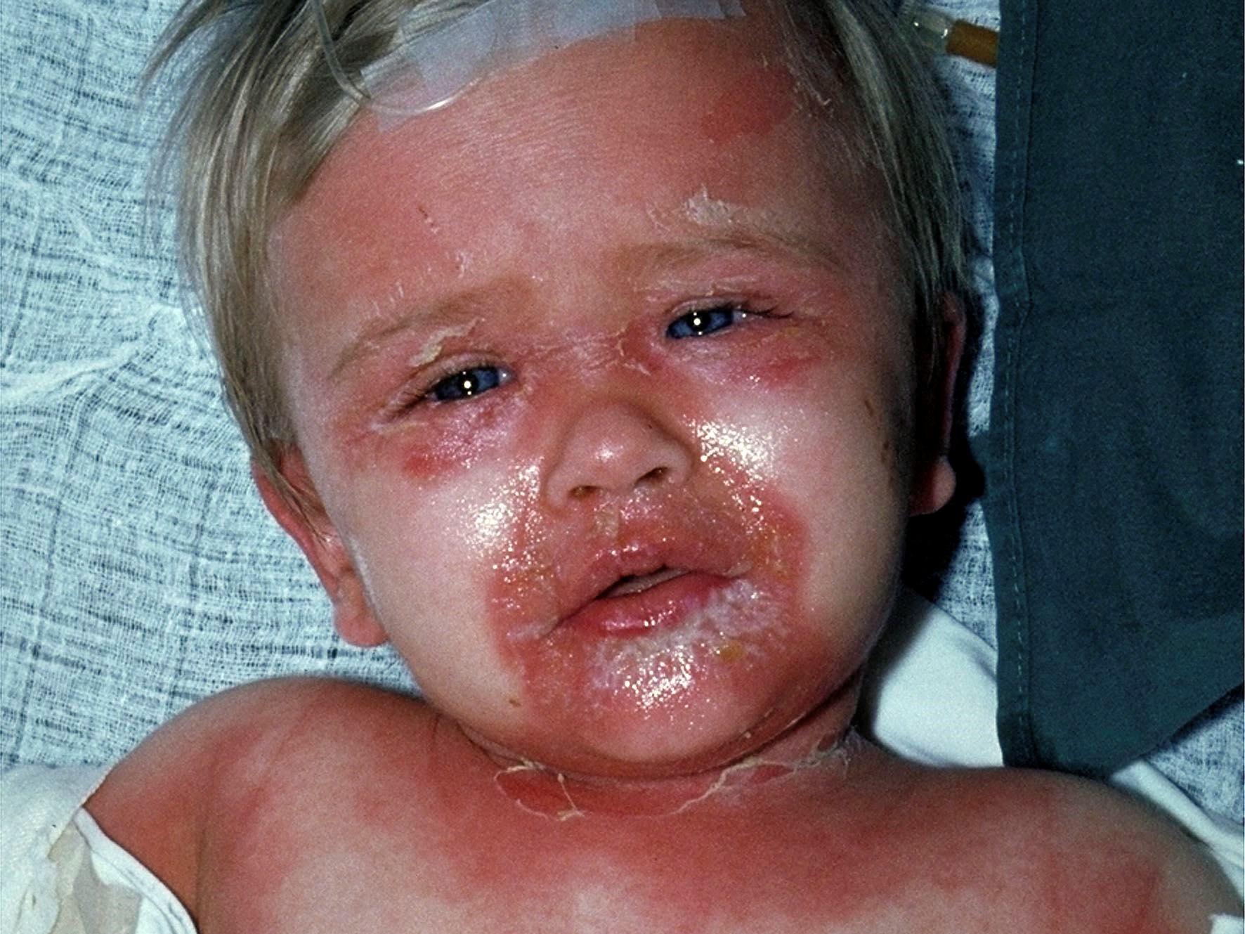
Illustration for DWII on SSSS
Image from DermNetNZ, provided by Prof. Raimo Suhonen.

What can possibly be new about SSSS? Four aspects – 1) epidemiology; 2) causation; 3) the clinical spectrum; and 4) therapeutic approach.

Epidemiology 

The incidence of SSSS seems to be increasing in recent years. The Nationwide Inpatient Sample 2008-2012 was analyzed, including a 20% sample of U.S. hospitalizations and 589 cases of SSSS. The mean annual incidence of SSSS was 7.67 per million U.S. children, with 45.1 cases per million U.S. infants age < 2 years. (2) Utilizing the same database, the annual incidence among U.S. adults was 0.98 cases per million. (3).

Causation 

To date, the exfoliative toxins have been associated with only certain strains of S. aureus, most of which have been methicillin-susceptible S. aureus (MSSA). As noted above, strains causing SSSS were usually reported as belonging to phage group II. More recent studies have reported isolates belonging to sequence types (ST) 15, ST121, ST 2126, and ST2993, using multilocus sequence typing (MLST). Of these, ST121 has been most commonly reported as a cause of SSSS. (4) Doudoulakakis et al had observed a sharp increase in SSSS settings since 2015, with 31 cases having been documented during the period 2014-2017. The molecular investigation of strains from that period demonstrated the emergence of a methicillin-susceptible, mupirocin- and fusidic acid-resistant Staphylococcus aureus clone belonging to the ST121 complex and carrying both epidermolysin (eta/etb) genes. (5) Additionally, while the SSSS exfoliation has been mainly attributed to the effects of the ETs, studies have also reported the Panton-Valentine leukocidin (PVL) genes associated with SSSS isolates. (4)

Clinical spectrum

Mazori et al note that SSSS classically presents with acute‐onset, generalized, tender erythema with flexural accentuation, flaccid blisters, periorificial crusting and radial fissuring; the Nikolsky sign is positive. As previously discussed, although the blisters of SSSS are sterile, S. aureus may be cultured from sites of infection or colonization where the toxins originate prior to dissemination, notably the conjunctivae, nares, perioral skin, axillae, umbilicus and anus. Patients are characteristically febrile, irritable, lethargic and anorexic. Although this classic severe phenotype is widely recognized in children, SSSS is spectrum of disease with mild and moderate variants. The authors presented four variant cases in infants. Physical examination demonstrated varying degrees of nontender erythema and superficial desquamation with periorificial, flexural and/or acral accentuation. All patients had a negative Nikolsky sign. One patient was febrile; vital signs were otherwise normal in all patients. One patient had leukocytosis. Although the reason for the SSSS phenotypic variability is unknown, it has been hypothesized that toxin load may play a role, with milder phenotypes having increased renal toxin clearance and/or higher levels of neutralizing antibodies. The authors emphasize that appreciating the variability of SSSS is crucial as the condition is potentially fatal; however, with medical management, the mortality rate in children is as low as 0.33%. (6)

SSSS in adults is a different story. Patients at risk include those who are immunosuppressed (notably HIV/AIDS), have severe renal impairment or failure, or in those with malignancies. The mortality rate is much higher in adults than children, having been reported as high as 59%. (7)

Therapeutic approach

Wang et al retrospectively studied 51 patients with SSSS; sensitivity data were available for 28 patients. Resistance to clindamycin was observed in 17 isolates (63%), erythromycin in 18 isolates (69%), and trimethoprim/sulfamethoxazole in 2 isolates (7%). No isolates were resistant to oxacillin or vancomycin. The authors concluded SSSS-associated isolates were more likely to be clindamycin-resistant and less likely to be methicillin-resistant compared to overall staphylococcal infections. They favor cephalosporins and penicillinase-resistant penicillins (eg, oxacillin) for empiric management of SSSS, with consideration of adding MRSA coverage in communities with high MRSA prevalence or failure to improve following several days of treatment. Although clindamycin has been touted to reduce toxin production, the authors found no benefit to adding clindamycin to the regimen. (8) 

Supportive care for management of dehydration, thermoregulation, and nutritional support is crucial. Emollients and non-adherent dressing should be applied to the skin and denuded areas to promote healing and reduce heat loss. (7)

In conclusion, even though it has been more than 140 years since it was first described, SSSS is a dynamic disorder with a spectrum of clinical presentations and an altered microbiology that demands our attention.

Point to remember: SSSS may present on a clinical spectrum and has been increasing in incidence. Although vastly more common in infants, adults with immunosuppression and renal impairment are at risk, with a much greater mortality. Changes in pathogenesis may be related to mupirocin resistance. 

Our expert's viewpoint

Bernard Cohen, MD
Professor of Dermatology and Pediatrics
Johns Hopkins University School of Medicine

There is clear evidence that the incidence of SSSS has increased dramatically in the United States over the last 2 decades. As a result, it is important that we train our medical colleagues, particularly pediatricians, to recognize the clinical findings and to distinguish SSSS from toxic epidermal necrolysis/Stevens-Johnson syndrome. They need to know that the toxins target desmoglein 1, which is present high in the epidermis, but not in mucous membranes.

Interestingly, in our series at the Johns Hopkins Children's Center none of the affected children had an identifiable primary bacterial infection; positive cultures were obtained from sites of colonization including the nares, conjunctivae, umbilicus, and perianal area. There was some variability in the clinical presentations, but most demonstrated classical findings including diffuse erythema, erosions around sites of minor trauma (particularly intertriginous and periorificial areas), and a positive Nikolsky sign. In our series we excluded 2 children who developed SSSS during prolonged hospitalizations for complications of prematurity. None of our patients were immunodeficient, on immunosuppressive medications, or had decreased renal function. 

Moreover, the length of hospitalization in our patients averaged 2 days, regardless of the antibiotic administered and whether or not the organism was sensitive or resistant to the antibiotic. Many clinicians have pushed for use of clinidamycin because it suppresses toxin production, but even in those patients who were given clindamycin montherapy, there was no change in length of hospitalization. Additionally, resistance of the causative organisms to clindamycin is approaching or exceeding 50%. 

It makes me wonder if an antibiotic is helpful when a primary infection is not identified in otherwise healthy immunocompetent patients. I understand that the standard of care is administration of systemic antibiotics, and in light of our finding that 100% were MSSA, we recommend a cephalosporin and penicillinase-resistant penicillins with consideration of adding MRSA coverage in communities with a high incidence of MRSA, until culture and sensitivities are available.

Management of pain, fluids and electrolytes, and good skin care is critical. We discouraged narcotics since pain usually decreased dramatically within 36-48 hours with aggressive use of topical emollients — non-stick dressings and debridement was a no-no! Frequent application of emollients to the crusty vermillion border was critical to maintain oral intake in all patients, including bottle and breast feeding in young infants.

In short, SSSS is not rare, hospitalization in otherwise healthy children is usually short with proper management, and primary infections in healthy children is uncommon. Therapeutically, use of antibiotics may not change the generally favorable outcome if good skin care is provided; however, in children and adults with immunosuppression and/or renal disease the risk of morbidity and mortality is significant, and these patients need to be evaluated and treated aggressively.

  1. Mockenhaupt M, Idzko M, Grosber M, Schöpf E, Norgauer J. Epidemiology of staphylococcal scalded skin syndrome in Germany. J Invest Dermatol 2005; 124: 700-703.

  2. Staiman A, Hsu DY, Silverberg JI. Epidemiology of staphylococcal scalded skin syndrome in US children. Br J Dermatol 2018; 178: 704-708. 

  3. Staiman A, Hsu DY, Silverberg JI. Epidemiology of staphylococcal scalded skin syndrome in US adults.

  4. Hultén KG, Kok M, King KE, Lamberth LB, Kaplan SL. Increasing number of Staphylococcal scalded skin syndrome caused by ST121 in Houston, Texas. Pediatr Infect Dis J 2020; 39: 30-34. 

  5. Doudoulakakis A, Spilopoulou I, Syridou G, Giormezis N, et al. Emergence of staphylococcal scalded skin syndrome associated with a new toxinogenic, methicillin-susceptible Staphylococcus aureus clone. J Med Micobiol 2019; 68: 48-51. 

  6. Mazori DR, Leonard A, Alexander JB, Glick SA. The spectrum of staphylococcal scalded skin syndrome: A case series in children. Br J Dermatol 2020; 45: 333-336. 

  7. Ross A, Shoff HW. Staphylococcal scalded skin syndrome. StatPearls [Internet], Treasure Island (FL). StatPearls Publishing; 2020 Jan. 

  8. Wang Z, Feig JL, Mannschreck DB, Cohen BA. Antibiotic sensitivity and clinical outcomes in staphylococcal scalded skin syndrome. Pediatr Dermatol 2020; 37: 222-223.


Sent from my iPhone

Benjamin Hidalgo-Matlock
Skin Care Physicians of Costa Rica

Clinica Victoria en San Pedro: 4000-1054
Momentum Escazu: 2101-9574

Please excuse the shortness of this message, as it has been sent from
a mobile device.
Thursday, April 22, 2021

Treatment of Patients Experiencing Dupilumab Facial Redness With Itraconazole and Fluconazole Journal of the American Academy of Dermatology

TAKE-HOME MESSAGE


Abstract

Dupilumab facial redness (DFR) is a recently described adverse event of dupilumab use.Prior cases of treatment with itraconazole have been reported.In this retrospective study, we investigated the management of DFR with itraconazole or fluconazole. This study was approved by the Tufts Medical Center (TMC) Institutional Review Board. Patients at TMC Dermatology who were on dupilumab for at least 16 weeks, had a diagnosis of DFR, and completed at least two weeks of daily fluconazole or itraconazole 200 milligrams (mg) were included. Investigator global assessment (IGA) before and after azole use, dupilumab associated ocular surface disease (DOSD), and self-reported improvement (%), obtained from a routine pharmacist phone call two weeks after azole initiation, were recorded. DOSD was defined as irritation, dryness, tearing, or conjunctivitis.

Journal of the American Academy of Dermatology
Treatment of Patients Experiencing Dupilumab Facial Redness With Itraconazole and Fluconazole: A Single Institutional Retrospective Medical Record Review
J Am Acad Dermatol 2021 Mar 25;[EPub Ahead of Print], DX Gao, S Song, JS Kahn, SR Cohen, K Fiumara, N Dumont, D Rosmarin 

Sent from my iPhone

Benjamin Hidalgo-Matlock
Skin Care Physicians of Costa Rica

Clinica Victoria en San Pedro: 4000-1054
Momentum Escazu: 2101-9574

Please excuse the shortness of this message, as it has been sent from
a mobile device.

Monday, April 19, 2021

Contact Allergy in Patients With Chronic Venous Leg Ulcers

TAKE-HOME MESSAGE

Abstract 

Contact allergy (CA) is prevalent in patients with chronic leg ulcers (CLUs) due to venous stasis1and may delay wound healing. Exposure to different ointments and wound dressings over time, combined with occlusive bandaging, may predispose to contact sensitization.2The spectrum of allergens depends on wound care practices.3, 4 Studies on CA in patients with CLUs from Norway are lacking. To determine the occurrence of CA in patients with CLUs, patch testing was performed with the Leg Ulcer Series (LUS) containing 27 chemicals and five additional substances relevant to wound treatment: Caine mix III and V, hydrocortisone, IntraSite gel (propylene glycol 20%, Smith & Nephew), and Brulidine (dibrompropamidine 0.15%, Sanofi‐Aventis) (Table S1). Fifty‐two of the 97 patients were also tested with the European Baseline Series (EBS) (Table S2).

Contact Dermatitis
Contact Allergy in Patients With Chronic Venous Leg Ulcers
Contact Derm 2020 Dec 27;[EPub Ahead of Print], AH Lossius, M Lorentzen, J Austad, TK Bergersen 


Sent from my iPhone

Benjamin Hidalgo-Matlock
Skin Care Physicians of Costa Rica

Clinica Victoria en San Pedro: 4000-1054
Momentum Escazu: 2101-9574

Please excuse the shortness of this message, as it has been sent from
a mobile device.

Sunday, April 18, 2021

Association Between Topical Calcineurin Inhibitor Use and Risk of Cancer JAMA Dermatology

TAKE-HOME MESSAGE


Abstract


IMPORTANCE

Topical calcineurin inhibitors (TCIs) are commonly used as second-line treatment for atopic dermatitis. In 2006, the US Food and Drug Administration issued a black box warning against TCI use, citing data from case reports and animal studies indicating a potential risk of cancer.

OBJECTIVE

To evaluate the association between TCI use and risk of malignant neoplasms compared with nonactive and active comparator groups.

DATA SOURCES

Electronic searches were conducted in MEDLINE via Ovid, Embase via Ovid, and Web of Science from database inception to August 21, 2020.

STUDY SELECTION

Observational studies investigating the association between treatment with TCIs (ie, tacrolimus and pimecrolimus) and the development of cancer with nonactive or active comparators were included. The population of interest was not limited to any specific disease state, age, or sex. All articles were assessed independently and in duplicate by 2 reviewers. Risk of bias was assessed using the Newcastle-Ottawa scale. Of 2464 nonduplicate records retrieved from the search, 11 studies met the inclusion criteria.

DATA EXTRACTION AND SYNTHESIS

Data extraction was conducted independently by 2 reviewers according to Preferred Reporting Items for Systematic Reviews and Meta-analyses (PRISMA) guidelines. Random-effects meta-analyses were used to derive pooled relative risk (RR) estimates. Data were analyzed from July 25 to October 25, 2020.

MAIN OUTCOMES AND MEASURES

Risk of cancer overall and risk of specific cancer types (lymphoma, melanoma, and keratinocyte carcinoma).

RESULTS

Eight unique cohort studies (408 366 treated participants [55.1% female], 1 764 313 nonactive comparator controls, and 1 067 280 controls using topical corticosteroids) and 3 unique case-control studies (3898 cases [55.0% male] and 14 026 cancer-free controls [52.4% male]) were included. There was no association between TCI use and cancer overall compared with nonactive comparators (RR, 1.03; 95% CI, 0.92-1.16). Lymphoma risk was elevated with TCI use with both nonactive (RR, 1.86; 95% CI, 1.39-2.49) and topical corticosteroid comparators (RR, 1.35; 95% CI, 1.13-1.61). No significant association was found between TCI use and increased skin cancer (melanoma and keratinocyte carcinoma).

CONCLUSIONS AND RELEVANCE

The findings of this systematic review and meta-analysis suggest an association between TCI use and risk of lymphoma but not other cancers. Combined with the low absolute risk of lymphoma, the potential increased risk attributable to TCI use for any individual patient is likely very small.


JAMA Dermatology
Association Between Topical Calcineurin Inhibitor Use and Risk of Cancer, Including Lymphoma, Keratinocyte Carcinoma, and Melanoma: A Systematic Review and Meta-Analysis
JAMA Dermatol 2021 Mar 31;[EPub Ahead of Print], M Lam, JW Zhu, M Tadrous, AM Drucker

Sent from my iPhone

Benjamin Hidalgo-Matlock
Skin Care Physicians of Costa Rica

Clinica Victoria en San Pedro: 4000-1054
Momentum Escazu: 2101-9574

Please excuse the shortness of this message, as it has been sent from
a mobile device.

Friday, April 16, 2021

Contact Sensitizations to Disinfectants Containing Alcohols or Quaternary Ammonium Compounds Contact Dermatitis


TAKE-HOME MESSAGE

Abstract
Contact Dermatitis
Contact Sensitizations to Disinfectants Containing Alcohols or Quaternary Ammonium Compounds Are Rarely of Clinical Relevance
Contact Derm 2021 Mar 24;[EPub Ahead of Print], K Kreipe, S Forkel, KE Heinemann, K Amschler, T Fuchs, J Geier, T Buhl

Nonavalent HPV Vaccine for the Treatment of Multiple Recalcitrant Warts Journal of the American Academy of Dermatology


TAKE-HOME MESSAGE

Abstract
Journal of the American Academy of Dermatology
Nonavalent Human Papilloma Virus Vaccine for the Treatment of Multiple Recalcitrant Warts: An Open Label Study
J Am Acad Dermatol 2021 Mar 27;[EPub Ahead of Print], JO Shin, JH Son, J Lee, HS Kim, HC Ko, BS Kim, MB Kim, K Shin

MTX en AA

Oral Methotrexate Monotherapy for Severe Alopecia Areata

TAKE-HOME MESSAGE

Abstract
Journal of cutaneous medicine and surgery
Oral Methotrexate Monotherapy for Severe Alopecia Areata: A Single Center Retrospective Case Series
J Cutan Med Surg 2021 Mar 09;[EPub Ahead of Print], M Kinoshita-Ise, M Sachdeva, SA Martinez-Cabriales, NH Shear, P Lansang

Laser en pelo junto con OC

Combined Oral Contraceptives or Metformin Plus Laser Treatment in Polycystic Ovarian Syndrome Hirsute Patients

Journal of Drugs in Dermatology

 1 Expert Comment

TAKE-HOME MESSAGE

abstract

This abstract is available on the publisher's site.

Topic Alerts

Click on any of these tags to subscribe to Topic Alerts. Once subscribed, you can get a single, daily email any time PracticeUpdate publishes content on the topics that interest you.

Visit your Preferences and Settings section to Manage All Topic Alerts


Journal of Drugs in Dermatology
Adding Combined Oral Contraceptives or Metformin to Laser Treatment in Polycystic Ovarian Syndrome Hirsute Patients
J Drugs Dermatol 2021 Mar 01;20(3)302-306, N Dorgham, A Sharobim, H Haggag, M El-Kalioby, D Dorgham