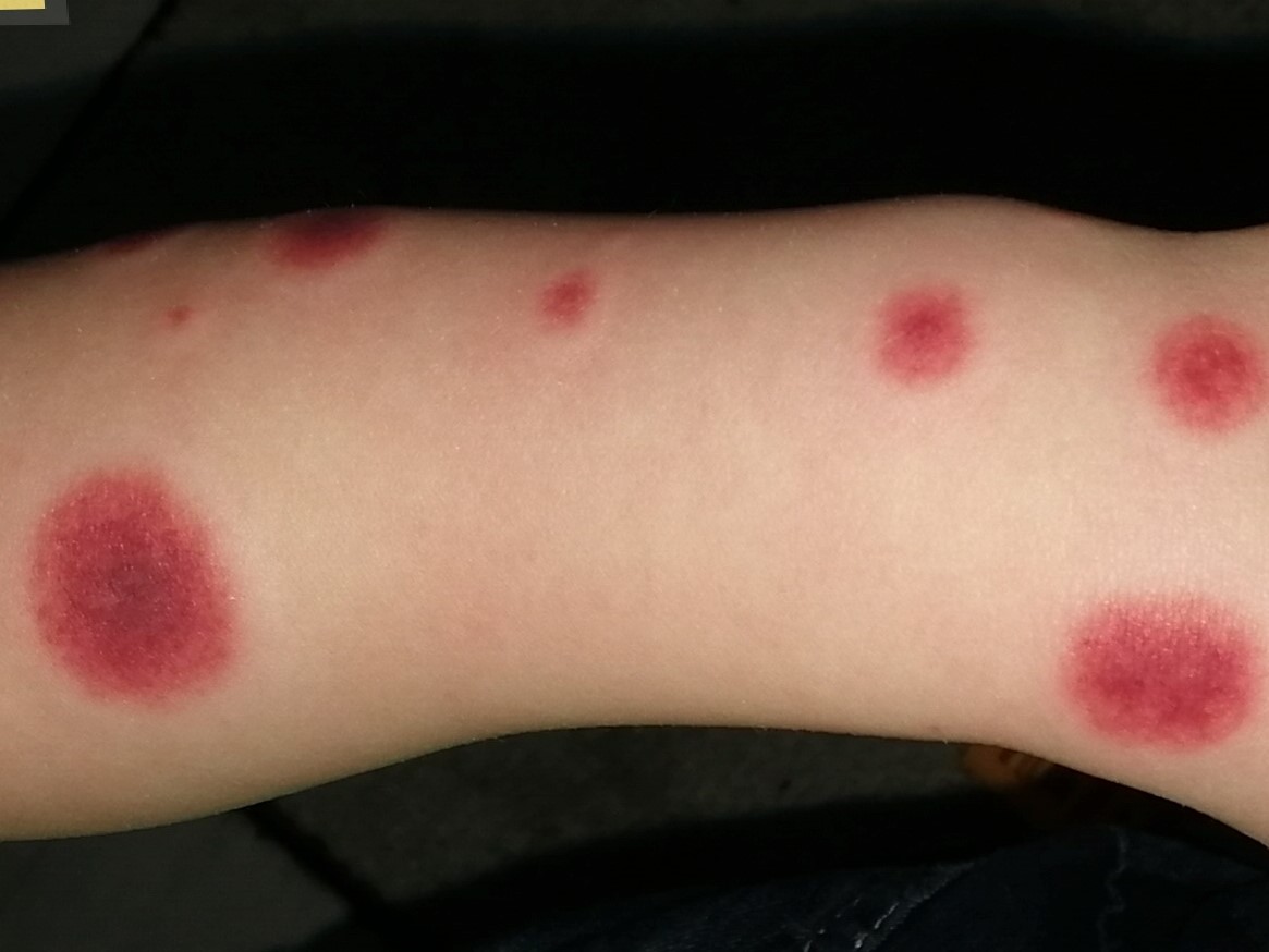
ALARMING BUT BENIGN: THE ENIGMA OF ACUTE HEMORRHAGIC EDEMA OF INFANCY

By Warren R. Heymann, MD, FAAD
Sept. 8, 2021
Vol. 3, No. 36

The quotation is from Irving Snow's 1913 publication which is considered the first description of acute hemorrhagic edema of infancy (AHEI, aka Finkelstein disease Seidlmayer syndrome, or post-infectious cockade purpura). AHEI is a rare, predominantly cutaneous leukocytoclastic vasculitis, typically diagnosed in children younger than 2 years of age, with a male: female ratio of 2 to 1. Varanaki et al are on point with this observation: "Its rapid onset and the dramatic character of skin lesions cause parental and also health care provider anxiety." (2,3) The diagnosis of AHEI is mostly based on clinical features with the following proposed criteria: 1) Age less than 2 years; 2) Purpuric or ecchymotic target-like lesions, with edema of the face, auricles, and extremities, with or without mucosal involvement; 3) Lack of systemic disease or visceral involvement; and 4) Spontaneous recovery within a few days or weeks. (4) Alarming presentations may include hemorrhagic lacrimation and epistaxis. Although visceral and systemic manifestations are uncommon compared to Henoch-Schönlein purpura, gastrointestinal bleeding, intussuception, and renal involvement have been reported. Auricular chondritis and epididymoorchitis have been observed. (3) Visual impairment due to massive periorbital edema and a compartment syndrome from foot edema have also been reported. (4)

Approximately 75% of cases of AHEI are preceded by respiratory infections, diarrheal illnesses, or urinary tract infections. Antibiotics and vaccinations have been identified as triggers [presumably inducing an immune complex-induced reaction]. Viruses including rotavirus, herpes simplex virus, and adenovirus have been implicated. Chesser et al reported the case of an 8-month-old girl with AHEI attributed to coronavirus NL63 by nucleic acid amplification testing. Her eruption cleared after a few days, but three weeks later, she had a brief recrudescence of the eruption. (5)
Although erythema multiforme, Kawasaki syndrome, Sweet syndrome, meningococcemia, and purpura fulminans all may be considered in the differential diagnosis, the main consideration is Henoch-Schönlein purpura (HSP). HSP is an acute IgA-mediated vasculitis involving the small vessels of the skin, the kidneys, the gastrointestinal tract, the joints, and, occasionally, the lungs and the central nervous system. It differs from AHEI by being observed in older patients (3-8 years) with purpuric lesions predominantly on the buttocks and legs, sparing the face and ears. HSP tends to persist for a longer time (>1 month). Systemic findings, notably abdominal pain, renal disease, and arthritis are common in HSP and can adversely affect the prognosis. (6)
Histologically, both AHEI and HSP demonstrate leukocytoclastic vasculitis, however, there are differences, most importantly in IgA deposition by direct immunofluorescence. HSP is strongly associated with IgA deposition; in AHEI, it is appreciated in approximately 25% of cases. (7) In their systematic review of 75 cases of AHEI (64 boys, 11 girls), Pellanda et al observed vessel wall deposition of complement C3 in 40 cases. IgM (N = 24), IgA (N = 21), IgG (N = 13), and IgE (N = 3) were less frequently detected. Gender, age, clinical features, and disease duration were not statistically different in cases with and without vessel wall deposition of IgA. Although the authors concluded that this data provides further evidence that AHEI and IgA vasculitis are different entities, they acknowledge that further studies are warranted to characterize the immunoglobulin A that is found in the vessel wall of some children with AHEI. (8)
Therapeutically, as AHEI is a self-limited disorder, usually no treatment is necessary. For those cases with significant edema, or rare systemic involvement, corticosteroids may hasten resolution. (7)
Over 100 years ago, Dr. Snow was perplexed about his patient. Although we now have a good sense of the nature and prognosis of AHEI and can reasonably reassure parents of the excellent prognosis — despite the dramatic presentation — questions remain. Is this a benign variant of HSP (at least in some cases) or a distinct entity? If it is an HSP variant, why do younger children fare better?
Point to Remember: Despite the dramatic presentation of acute hemorrhagic edema of infancy, the majority of patients have an excellent prognosis. Differentiation from Henoch-Schönlein purpura by history, physical examination, laboratory findings, and (if deemed necessary) histopathology will guide management.
Our expert's viewpoint
Anthony J. Mancini, MD
Professor of Pediatrics & Dermatology
Ann & Robert H. Lurie Children's Hospital of Chicago
Northwestern University Feinberg School of Medicine
Purpura in a child often triggers clinical alarm, and rightfully so, as many of the potential causes entail a risk for internal organ involvement and significant morbidity (and, in some cases, mortality). Acute hemorrhagic edema (AHE) is notable for an ominous presentation which is disproportionate to the overall-well status of the infant or young toddler. Some useful distinguishing features which may point the clinician in the right direction include the "cockade" (or medallion-like) cutaneous lesions with scalloped borders and annularity; centrifugal progression and expansion of the skin lesions from edematous papules and petechiae, with eventual coalescence; and edema of the face, distal extremities, and occasionally the scrotum or penis in males. Although serious internal involvement is extremely rare as pointed out by Dr. Heymann, a significant number of patients had some systemic symptoms in one large series, including arthralgias, arthritis, gastrointestinal hemorrhage, or microscopic hematuria. Overall, though, the typical course for AHE is marked by rapid onset, a short benign course, and complete recovery, typically over one to three weeks. While the debate over whether AHE represents a variant of Henoch-Schönlein purpura versus a distinct entity continues, the usual lack of internal manifestations and IgA immune deposits, as well as the benign course with no propensity toward recurrence, tips the scale in favor of the latter.
Dr. Mancini had disclosed financial relationships with the following to the AAD at the time of publication: Cassiopia, Dermavant Sciences, Inc., and Pfizer Inc. Full disclosure information is available at coi.aad.org.
Snow IM. Purpura, urticaria and angioneurotic edema of the hands and feet in a nursing boy.
Varanaki ME, Ladomenou F, Anatoliltaki M, Vlachaki G. Review and case report of acute hemorrhagic edema of infancy. A benign cause of a striking rash. Eur J Pediatr Dermatol 2019; 29: 134-138.
Mreish S, Al-Tatari H. Hemorrhagic lacrimation and epistaxis in acute hemorrhagic edema of infancy. Case Rep Pediatr 2016;2016:9762185.
Chavez-Alvarez S, Barbosa-Moreno L, Ocampo-Garza J, Ocampo-Candiani. Acute hemorrhagic edema of infancy (Finkelstein's disease): favorable outcome with systemic steroids in a female patient. An Bras Dermatol 2017; 92: 150-152.
Chesser H, Chambliss JM, Zwemer E. Acute hemorrhagic edema of infancy after coronavirus infection with recurrent rash. Case Rep Pediatr 2017;2017:5637503.
Miconi F, Cassiani L Savaresse E, Celi F, et al. Targetoid skin lesions in a child: Acute hemorrhagic oedema of infancy and its differential diagnosis. Int J Environ Res Public Health 2019; 16: 823 doi: 10.3390/ijerph16050823.
Rohr, Manalo IF, Mowad C. Acute hemorrhagic edema of infancy: Guide to prevent misdiagnosis. Cutis 2018; 102: 359-362.
Pellanda G, Lava SAG, Miliani GP, Bianchetti MG. Immune deposits in skin vessels of patients with acute hemorrhagic edema of young children: A systematic literature review. Pediatr Dermatol 2019; Nov 22 [Epub ahead of print]
Skin Care Physicians of Costa Rica
Clinica Victoria en San Pedro: 4000-1054
Momentum Escazu: 2101-9574
Please excuse the shortness of this message, as it has been sent from
a mobile device.
posted by dermatica at
September 08, 2021
![]()
![]()

0 Comments:
Post a Comment
Subscribe to Post Comments [Atom]
<< Home