Dermatología en Costa Rica

Saturday, April 30, 2022

Published in Dermatology News · April 29, 2022 Annual Costs Estimated for Wart Treatment in the U.S. for 2015-2017

Per-patient costs for evaluation and management estimated at $288.28 for cutaneous warts and $431.47 for anogenital warts in 2019


Research Letter 
April 27, 2022

Annual Health Care Utilization and Costs for Treatment of Cutaneous and Anogenital Warts Among a Commercially Insured Population in the US, 2017-2019

JAMA Dermatol. Published online April 27, 2022. doi:10.1001/jamadermatol.2022.0964

Cutaneous warts are common growths responsible for several million office visits per year.1 While multiple treatments are available, including topical treatments, intralesional immunotherapy, and cryotherapy,2,3 little is known about health care utilization and costs associated with different treatments.


Sent from my iPhone

Benjamin Hidalgo-Matlock
Skin Care Physicians of Costa Rica

Clinica Victoria en San Pedro: 4000-1054
Momentum Escazu: 2101-9574

Please excuse the shortness of this message, as it has been sent from
a mobile device.

Thursday, April 28, 2022

Oximetazolina para ptosis palpebral

EYE-OPENING NEWS ABOUT OXYMETAZOLINE


DII small banner

By Warren R. Heymann, MD, FAAD
April 27, 2022
Vol. 4, No. 17

Certain articles have a "wow" factor that immediately impacts practice. In 2007, I recall being impressed by the 2 cases of erythematotelangiectatic rosacea reported by Shanler and Ondo that dramatically improved with the application of oxymetazoline hydrochloride 0.05%. (1) Oxymetazoline had been used for decades to treat nasal decongestion or to "get the red out" of the eyes; I thought the idea was brilliant. I have been recommending it ever since either as the over-the-counter product or the 1% prescription cream formulation released in 2017. (2)

Oxymetazoline is a potent vasoconstrictor that also demonstrates anti-inflammatory properties. It is a direct-acting, imidazoline-type sympathomimetic agonist that is highly selective for the α1A-adrenoceptor and is also a partially selective α2A-receptor agonist. (1) The drug's vasoconstrictive effects have been used to advantage for other dermatological conditions, including post-acne erythema (3,4), erythromelalgia of the knee (5), and hemostasis during dermatologic surgery. (6) 

Oxymetazoline 0.1% ophthalmic solution is now available for the treatment of blepharoptosis (drooping of the eyelid, subsequently referred to as ptosis). This commentary will focus on the use of oxymetazoline for ptosis. (I have no financial or any other perceived conflict of interest with RVL Pharmaceuticals, Inc. the distributor of Upneeq.)

Ptosis may be a cosmetic concern but if significant enough, may interfere with vision. I have mild ptosis — people have told me to "wake up" during our conversations. (Sometimes they are correct and I am falling asleep!)

Ptosis is a physical sign, not a diagnosis. Ptosis may be congenital or acquired. Acquired adult ptosis is categorized as 1) aponeurotic (aka involutional; this is the most common form, usually presenting in the 5th-6th decade, most frequently due to dehiscence or disinsertion of the levator aponeurosis; 2) neurogenic (due to disruption of innervation of either the levator muscle or Muller muscle [a small smooth muscle arising from the striated levator muscle along with the aponeurosis] as may be seen with Horner syndrome or 3rd cranial nerve palsy; 3) myogenic (due to an abnormality of levator muscle itself, as seen with myasthenia gravis or myotonic dystrophy; 4) mechanical (secondary to any tumor causing increased eyelid weight, conjunctival scarring, and blepharochalasis); and 5) traumatic (due to direct or indirect injury to the levator muscle). (7) If there is any question about the etiology of ptosis an ophthalmological consultation should be sought. 

Oxymetazoline elevates the eyelid by activating α-adrenergic receptors in the Muller muscle. Slonim et al reported the results of 2 industry-sponsored randomized placebo-controlled masked trials that assessed the efficacy of oxymetazoline, 0.1%, eyedrops for ptosis. A total of 304 patients with acquired ptosis to topical oxymetazoline HCl, 0.1%, vs vehicle in 2 trials were randomized. All participants had clinically relevant involutional (aponeurotic) ptosis defined by superior visual field loss and reduced eyelid height. Response to therapy was evaluated at 1 day and 14 days of treatment, and safety was evaluated over 42 days of follow-up. Using Leicester Humphrey Visual Field (HVF) static perimetry as the primary outcome, the authors showed a change in the oxymetazoline treatment arm of 4 to 5 points of superior visual field more than the change in the vehicle arm. The secondary outcome, change in upper eyelid height, increased by less than 0.5 mm in the treatment arm compared with the vehicle arm on day 1 and by 0.67 mm compared with vehicle on day 14.

Oxymetazoline HCl, 0.1%, was generally well tolerated, with no change in visual acuity or intraocular pressure but with somewhat higher rates of punctate keratitis, conjunctival hyperemia, and subjectively blurred vision in the study group. (8,9) In an editorial accompanying the article by Slonim at al, Bradley and Bradly opine; "It is unclear whether a gain of 4 to 5 points of superior visual field, or roughly half a line of points on the Leicester HVF, represents meaningful clinical change. The corresponding less than 1 mm of eyelid elevation produced by oxymetazoline compared with vehicle may also be of marginal clinical benefit. While it is possible that this degree of eyelid elevation yields substantial improvements in visual function and patient self-image, an assessment of health-related quality of life was not included in the study." Additionally, this study did not compare oxymetazoline with surgery, although when compared to historical data, the improvement with oxymetazoline is less than is usually observed with surgical repair. (9)

Wirta et al (with Slonim as senior author) evaluated the safety profile of oxymetazoline 0.1% when administered once daily for 14-84 days to assess treatment-emergent adverse events (TEAE). A total of 568 participants with acquired blepharoptosis were evaluated. The median age was 66 years and 74.8% of participants were female. Overall, 375 participants self-administered oxymetazoline 0.1% to both eyes once/day and 193 self-administered placebo (vehicle) daily. TEAE incidence was similar among participants using oxymetazoline 0.1% (31.2%) or vehicle (30.6%). Nearly all TEAEs were mild-to-moderate, and most were not suspected of being treatment related. Serious TEAEs occurred in four participants receiving oxymetazoline 0.1% and one participant receiving vehicle. Nine and 2 participants in the oxymetazoline 0.1% and vehicle groups, respectively, discontinued due to a TEAE. Ocular TEAEs occurring in ≥2% of participants receiving oxymetazoline 0.1% were punctate keratitis, conjunctival hyperemia, dry eye, blurred vision, instillation site pain, and corneal vital dye staining, with none occurring in >3.5% of participants, allowing the authors to conclude that oxymetazoline for ptosis is safe and well tolerated.

Wirta et al explain that oxymetazoline may affect α-adrenergic receptors expressed elsewhere in the eye (conjunctiva, iris-ciliary structures, aqueous outflow tract) that could explain the adverse reactions of punctate keratitis, conjunctival hyperemia, and dry eye. Finally, oxymetazoline may be associated with transient mydriasis and acute angle closure glaucoma – trials with oxymetazoline excluded such patients. (10)

Dermatologists may encounter ptosis due to localized spread of botulinum toxin when administered for treating glabellar lines. Oxymetazoline may be considered as convenient, rapid, safe, and effective treatment for this adverse reaction to botulinum toxin. (11)

Before prescribing oxymetazoline, I would encourage all to read the package insert. Avoid using the medication in those with a history of acute angle glaucoma and be judicious with its use in hypertensive patients. 

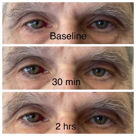
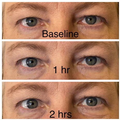
I could not resist the temptation. I tried it, but my results were barely perceptible (see below, and please ignore the subconjunctival hemorrhage). Our practice manager, however, was delighted with her results that were observed within an hour (see below, with permission).

Acknowledgement: I thank Lacy L. Sommer, MD, FAAD, for her photographic skills. 

Warren R. Heymann, MD, FAAD 
Image for DWII on ptosis

Practice manager
Image for DWII on ptosis

Point to Remember: Oxymetazoline has been used increasingly and successfully for its vasoconstrictive properties in rosacea, erythromlelagia, and post-acne erythema. The α-adrenergic receptors in the Muller muscle allow the drug to benefit patients with ptosis. 

Our expert's viewpoint

Mark S. Nestor, MD, PhD, FAAD
Director, Center for Cosmetic Enhancement
Director, Center for Clinical and Cosmetic Research
Aventura, FL
Voluntary Professor
Department of Dermatology and Cutaneous Surgery
Department of Surgery, Division of Plastic Surgery
University of Miami Miller School of Medicine
Miami, FL

While uncommon and ultimately self-limiting, botulinum toxin A induced blepharoptosis can cause distress both for the patient and for the treating physician. It occurs due to a toxin-induced weakness of the levator palpebrae superioris muscle. It normally is seen between three and 14 days after injection and normally resolves as the effect of the toxin wanes. While it is most commonly caused by inappropriate injection in high-risk zones it also can occur based on anatomical differences in some patients whereby a supraorbital foramen maybe present which causes a shortcut from the brow area to the muscle. Risk definitely decreases with experience and thorough anatomic knowledge of risk areas. A new treatment option (Oxymetazoline HCL 0.1%, Upneeq, RVL Pharmaceuticals) is now available which can significantly reduce the intensity and duration of the induced ptosis. (11)

Dr. Nestor had disclosed financial relationships with the following to the AAD at the time of publication: Aerolase, Allergan, Inc, Arcutis Biotherapeutics, Biofrontera AG, Bioregenerative Aesthetics, Inc., BirchBioMed, Brickell Biotech, Inc., Cara Therapeutics, Castle Biosciences, Inc, Croma-Pharma, Dermata Therapeutics, DermTech Inc., DUSA Pharmaceuticals, Eirion Therapeutics, Fastox Pharma, Ferndale Laboratories, Inc., Galderma Laboratories, LP, GmbH Austria, Ipsen, Kimera Labs, Inc, Krystal Biotech, Inc, MediWound, Merz Pharmaceuticals, LLC, Novaestiq Corp, Pulse Biosciences, Revian LTD., Rohrer Aesthetics, Seaspire Skincare, Sensus Healthcare, Sirnaomics, Inc., SkinJect Inc., Strathspey Crown, Suneva Medical, Inc., Vial. Full disclosure information is available at coi.aad.org.

  1. Shanler SD, Ondo AL. Successful treatment of the erythema and flushing of rosacea using a topically applied selective alpha1-adrenergic receptor agonist, oxymetazoline. Arch Dermatol. 2007 Nov;143(11):1369-71. doi: 10.1001/archderm.143.11.1369. PMID: 18025359.

  2. Okwundu N, Cline A, Feldman SR. Difference in vasoconstrictors: oxymetazoline vs. brimonidine. J Dermatolog Treat. 2021 Mar;32(2):137-143. doi: 10.1080/09546634.2019.1639606. Epub 2019 Aug 12. PMID: 31294643.

  3. Kalantari Y, Dadkhahfar S, Etesami I. Post-acne erythema treatment: A systematic review of the literature. J Cosmet Dermatol. 2022 Apr;21(4):1379-1392. doi: 10.1111/jocd.14804. Epub 2022 Feb 3. PMID: 35076997.

  4. Agamia N, Essawy M, Kassem A. Successful treatment of the face post acne erythema using a topically applied selective alpha 1-Adrenergic receptor agonist, oxymetazoline 1.5%, a controlled left to right face comparative trial. J Dermatolog Treat. 2022 Mar;33(2):904-909. doi: 10.1080/09546634.2020.1789045. Epub 2020 Jul 7. PMID: 32602755.

  5. Altemir A, Iglesias-Sancho M, Barrabés-Torrella C, Sanchez-Regaña M, Salleras-Redonnet M. Successful treatment of knee erythromelalgia with topical oxymetazoline. Clin Exp Dermatol. 2022 Apr;47(4):778-780. doi: 10.1111/ced.15060. Epub 2022 Jan 4. PMID: 34905256.

  6. Barklund JS, Wong EB, Brown M. Use of Oxymetazoline Hydrochloride 0.05% Soaked Pledgets for Hemostasis of Exposed Nasal Mucosa After Dermatologic Surgery. Dermatol Surg. 2021 Feb 1;47(2):305-306. doi: 10.1097/DSS.0000000000002272. PMID: 31809355.

  7. Koka K, Patel BC. Ptosis Correction. 2021 Nov 2. In: StatPearls [Internet]. Treasure Island (FL): StatPearls Publishing; 2022 Jan–. PMID: 30969650.

  8. Slonim CB, Foster S, Jaros M, Kannarr SR, Korenfeld MS, Smyth-Medina R, Wirta DL. Association of Oxymetazoline Hydrochloride, 0.1%, Solution Administration With Visual Field in Acquired Ptosis: A Pooled Analysis of 2 Randomized Clinical Trials. JAMA Ophthalmol. 2020 Nov 1;138(11):1168-1175. doi: 10.1001/jamaophthalmol.2020.3812. PMID: 33001144; PMCID: PMC7530825.

  9. Bradley EA, Bradley DJ. Oxymetazoline for Ptosis. JAMA Ophthalmol. 2020 Nov 1;138(11):1176-1177. doi: 10.1001/jamaophthalmol.2020.3833. PMID: 33001145.

  10. Wirta DL, Korenfeld MS, Foster S, Smyth-Medina R, Bacharach J, Kannarr SR, Jaros MJ, Slonim CB. Safety of Once-Daily Oxymetazoline HCl Ophthalmic Solution, 0.1% in Patients with Acquired Blepharoptosis: Results from Four Randomized, Double-Masked Clinical Trials. Clin Ophthalmol. 2021 Oct 8;15:4035-4048. doi: 10.2147/OPTH.S322326. PMID: 34675472; PMCID: PMC8517985.

  11. Nestor MS, Han H, Gade A, Fischer D, Saban Y, Polselli R. Botulinum toxin-induced blepharoptosis: Anatomy, etiology, prevention, and therapeutic options. J Cosmet Dermatol. 2021 Oct;20(10):3133-3146. doi: 10.1111/jocd.14361. Epub 2021 Aug 11. PMID: 34378298.


Sent from my iPhone

Benjamin Hidalgo-Matlock
Skin Care Physicians of Costa Rica

Clinica Victoria en San Pedro: 4000-1054
Momentum Escazu: 2101-9574

Please excuse the shortness of this message, as it has been sent from
a mobile device.

Monday, April 25, 2022

Journal Scan / Research · April 20, 2022 Leukocyte Adhesion and Rolling in Skin With Patient Outcomes After Hematopoietic Cell Transplantation Using Noninvasive Reflectance Confocal Videomicroscopy JAMA Dermatology


TAKE-HOME MESSAGE


Abstract 

IMPORTANCE

Hematopoietic cell transplantation (HCT) is a potential cure for hematologic cancer but is associated with a risk of relapse and death. Dynamic biomarkers to predict relapse and inform treatment decisions after HCT are a major unmet clinical need.

OBJECTIVE

To identify a quantitative characteristic of leukocyte-endothelial interactions after HCT and test its associations with patient outcomes.

DESIGN, SETTING, AND PARTICIPANTS

In this prospective single-center cohort study from June 2017 to January 2020, patients of any age, sex, race, and ethnicity who had HCT for hematologic cancer were referred by health care professionals as either suspected of having symptoms or not having symptoms of acute graft-vs-host disease between 25 and 161 days after HCT. Patients underwent noninvasive skin videomicroscopy. Videos of dermal microvascular flow were recorded with a reflectance confocal microscope. Two blinded observers (J.R.P. and Z.Z.) counted leukocytes adherent to and rolling along the vessel wall per hour (A&R). Of 57 enrolled patients, 1 relapsed before imaging and was excluded, resulting in 56 patients included in analyses.

MAIN OUTCOMES AND MEASURES

Relapse of cancer, relapse-free survival, and overall survival.

RESULTS

Among the 56 patients (median age, 59 years; 38 [68%] male) who underwent imaging a median of 40 days after HCT, 21 had high A&R and 35 had low A&R. After correcting for the revised Disease Risk Index, patients with high A&R had higher rates of relapse (hazard ratio [HR], 4.24; 95% CI, 1.32-13.58; P = .02), reduced relapse-free survival (HR, 3.29; 95% CI, 1.26-8.55; P = .02), and reduced overall survival (HR, 3.06, 95% CI, 1.02-9.19; P = .05). These associations were preserved after correcting for possible confounders, steroid treatment, and acute graft-vs-host disease status. In the prognostic adequacy calculation by using Cox models, the new imaging biomarker (A&R) accounted for 82% to 95% of the prognostic information to predict each outcome. By contrast, the best existing clinical predictor routinely available, the revised Disease Risk Index, accounted for 10% to 28% of the prognostic information in the same model.

CONCLUSIONS AND RELEVANCE

In this cohort study, leukocyte-endothelial interactions, visualized directly in skin after HCT, were associated with the patient outcomes of relapse, relapse-free survival, and overall survival. Assessing this dynamic marker could help patients at high risk for relapse who may benefit from interventions, such as early withdrawal of immunosuppression.


JAMA Dermatology
Association of Leukocyte Adhesion and Rolling in Skin With Patient Outcomes After Hematopoietic Cell Transplantation Using Noninvasive Reflectance Confocal Videomicroscopy
JAMA Dermatol 2022 Mar 26;[EPub Ahead of Print], I Saknite, JR Patrinely, Z Zhao, H Chen, A Beeghly-Fadiel, TK Kim, M Jagasia, M Byrne, ER Tkaczyk 

Sent from my iPhone

Benjamin Hidalgo-Matlock
Skin Care Physicians of Costa Rica

Clinica Victoria en San Pedro: 4000-1054
Momentum Escazu: 2101-9574

Please excuse the shortness of this message, as it has been sent from
a mobile device.
Journal Scan / Research · April 22, 2022 Disproportional Signal of Sexual Dysfunction Associated With Finasteride Use in Young Men With Androgenetic Alopecia Journal of the American Academy of Dermatology

TAKE-HOME MESSAGE


Abstract 

In a recent study by our group, finasteride use was found to be associated with increased reporting of suicidality and depression in young patients with androgenetic alopecia. This previous analysis suggested that the association between finasteride and depression might be mediated by sexual dysfunction, which in men includes erectile dysfunction, diminished libido, and ejaculatory disorders. It is challenging to characterize the association between finasteride use and sexual dysfunction at the population level due to underlying age-related comorbidities causing sexual dysfunction in older men with benign prostatic hyperplasia (BPH) and possible nocebo phenomenon in which a negative outcome occurs due to expectations that an intervention will cause harm, amongst other factors.

Journal of the American Academy of Dermatology
Disproportional signal of sexual dysfunction reports associated with finasteride use in young men with androgenetic alopecia: A pharmacovigilance analysis of VigiBase
J Am Acad Dermatol 2022 Mar 26;[EPub Ahead of Print], DD Nguyen, P Herzog, EB Cone, M Labban, KC Zorn, B Chughtai, S Basaria, DS Elterman, QD Trinh, N Bhojani 

Sent from my iPhone

Benjamin Hidalgo-Matlock
Skin Care Physicians of Costa Rica

Clinica Victoria en San Pedro: 4000-1054
Momentum Escazu: 2101-9574

Please excuse the shortness of this message, as it has been sent from
a mobile device.
Wednesday, April 20, 2022

ONE TOKER OVER THE LINE: CLARIFYING TOKER CELL HYPERPLASIA IN ZUSKA DISEASE AND ITS RELATIONSHIP TO HIDRADENITIS SUPPURATIVA

DII small banner

By Warren R. Heymann, MD, FAAD
April 20, 2022
Vol. 4, No.

Don't fret. I had never heard of Zuska disease (ZD) either until I read the article by Torre-Castro et al discussing the "tricky association" of Toker cell hyperplasia (TCH) in ZD. (1) Why should you care? Because it is likely that you have seen — and will continue to see — patients with this disorder and should be aware of this potentially problematic association.

ZD (aka mammillary fistula [MF]) was first reported by Zuska et al in 1951 who described a case series of 5 women (with his wife as the index patient) in what were previously considered nontuberculous breast fistulae. The presumption was that lactiferous ducts became obstructed by squamous metaplasia, resulting in subareolar breast abscesses. The mean age of presentation of ZD is 47 years and smoking is strongly associated with the disease. A rare complication of ZD is squamous cell carcinoma of the breast. (2,3) 

Cosman and Al-Refaie were the first to recognize extramammary cutaneous findings in ZD, reporting on 2 women in their forties, with evidence of hidradenitis suppurativa (HS) in the inguinal and axillary regions, respectively. The following are the authors' conclusions: "We present two patients with MF and concurrent acne inversa/HS lesions. These cases raise the question of whether MF might be a manifestation of acne inversa. Despite the ample literature on MF and the very extensive literature on HS, this is a previously unrecognized association, suggesting either that these cases are unusual or that they demonstrate something that has not been seen because it has not been looked for. In its lack of description of associated conditions, the literature contains the implicit assumption that MF is an isolated lesion of the breast. Our cases challenge that assumption, and it remains for surgeons who see MF frequently to confirm or deny the suggested association with acne inversa/HS." (4)

Toker cells (TC) are normal components of skin of the nipple and areola, thought to be derived from epidermally located mammary ductal epithelium. TC are observed in approximately 10% of nipple sections with routine stains and up to 90% of nipple specimens by CK7 staining, with greater numbers of cells located around nipple orifices. TC may be seen as isolated cells, in small clusters, or as gland-like structures. TCH refers to cases in which TC are found in increased numbers (defined as up to 20 single cells or up to 10 glands). The practical issue is in differentiating TCH from it mimic — Paget disease (PD) of the nipple. Paget cells tend to be more pleomorpbic and mitotic figures may be observed, compared to TC which, at most, have mild cytological atypia. In problematic cases, histochemical stains for mucin or immunostains may be needed to differentiate the two. (Paget cells stain with mucin stains such as mucicarmine, while TC are negative. Immunohistochemically, TC stain for CK7, CAM5.2, and EMA; variable staining is seen for ER and PR, and do not show significant staining for HER2, CK20, GCDFP-15, S100 protein, CD138, p53,p63, or CEA. Paget cells similarly stain with CK7, CAM5.2 and EMA and also show variable staining for ER and PR. Most importantly, the vast majority of cases of Paget disease are strongly HER2 positive; this may be considered the most helpful immunostain differentiating PD from TCH.) (5) 

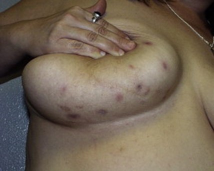
DWII image on Zuska disease
JAAD 2009; 60: 539-561.

Torre-Castro presented the case of a 42 year-old smoker with recurrent painful inflammatory lesions of her nipples, presenting as nipple inversion. Biopsies were performed because of the clinical concern for PD. The presence of large pale cells was initially thought to be consistent with PD — further differentiating immunohistochemical stains were performed confirming the correct diagnosis of TCH when the patient provided additional history of having similar inflammatory lesions of her groin and having a sister with HS. (1)

Squamous cell carcinoma (SCC) may be considered the most severe complication of HS — I am still haunted by my examination of a woman I encountered as a first-year dermatology resident. She had a fungating, filiform anal lesion, emanating from her scarred perianal HS, that tracked up to her cervical spine, ultimately causing her demise. The prevalence of SCC associated with HS is approximately 4.6%* and is more common among men. (*SCC complicating HS is considered rare — the reported prevalence seems high in my estimation.) As in other scars that predispose to the development of SCC, presumably chronic irritation and inflammation of HS drive the malignant transformation to SCC; additionally, other potential risk factors for malignant transformation may include human papillomavirus (HPV) infection and tobacco use. (6) I surmise that the cases of SCC of the breast in ZD are essentially the same process. 

In my PubMed search (performed Jan. 1, 2021) I cannot find any cases of PD in patients with ZD or HS. Of course, anything can happen. Torre-Castro et al have done dermatologists a service by pointing out how we need to be cognizant of TCH, so patients do not get misdiagnosed as PD in the context of ZD/HS. Such patients have enough to deal with already. 

Point to Remember: In all likelihood, Zuska disease is a localized form of hidradenitis suppurativa. Although this may rarely be complicated by squamous cell carcinoma, the development of Paget Disease is yet to be reported. Should Pagetoid cells be observed histologically, Toker cell hyperplasia — a benign condition — must be considered. Mucin and immunohistochemical stains will differentiate the two. 

Our expert's viewpoint

Ginette A. Okoye, MD, FAAD
Professor & Chair
Howard University College of Medicine

Mammillary fistula (MF) is likely a manifestation of hidradenitis suppurativa (HS). The demographics, anatomic location, and association with smoking and squamous cell carcinoma (SCC) support this assertion. Although submammary abscesses and sinus tracts are common in HS, involvement of the mammary tissue, especially in the sub-areola area, is less common and should be alarming to the patient and provider. I have several patients with HS and mamillary fistulae. These lesions are not subtle: my patients have sub-areola nodules and/or induration, inverted nipples, overlying peau d'orange, and purulent drainage from the nipple or areola. I refer these patients for urgent mammograms, and I send them to a breast surgeon for excision, if needed. Unfortunately, recurrence is common after excision, especially if the patient is not on concurrent anti-inflammatory therapy. Additionally, the excisions often result in scarring and asymmetry of the breasts, and exacerbation of the dysmorphia that plagues patients living with HS. My ongoing concern is that we could miss a diagnosis of breast cancer in patients with MF given their distorted anatomy, scar tissue, and blunting of our response to the recurrent 'false alarms' seen on their breast exams.

Additionally, Dr. Heymann's commentary highlights the gap in our understanding of the histopathology of hidradenitis suppurativa. Some of the histologic features of HS overlap with SCC (7) and (apparently!) Paget's disease, so building our expertise in the histopathology of HS has important implications. Patients with HS are increasingly undergoing en bloc excisions and de-roofing procedures. I strongly advocate for IRB-approved prospective collection of the discarded tissue from these procedures to help build HS tissue biorepositories. The availability of this tissue (and the associated clinical information) will facilitate observations and research to improve our understanding of this terrible disease. (8)

  1. Torre-Castro J, Haya-Martínez L, Ruffin-Vicente B, Moya-Martínez C, Núñez-Hipólito L, Díaz de la Pinta J, Cullen-Aravena D, Jo-Velasco M, Requena L. Toker cell hyperplasia in Zuska disease: A tricky association. J Cutan Pathol. 2021 Jan;48(1):180-183.

  2. Zuska JJ, Crile G Jr, Ayres WW. Fistulas of lactifierous ducts. Am J Surg. 1951 Mar;81(3):312-7. 

  3. Huws AM, Semkin L, Moalla A, Udayasankar S, Holt SDH, Sharaiha YM. Primary squamous cell carcinoma of the breast in association with Zuska's disease. Breast Cancer. 2018 May;25(3):365-369. 

  4. Cosman BC, Al-Refaie WB. Mammillary fistula as a manifestation of acne inversa (hidradenitis suppurativa): report of two cases. J Am Coll Surg. 2002 Jun;194(6):829-33. 

  5. Torous VF, Schnitt SJ, Collins LC. Benign breast lesions that mimic malignancy. Pathology. 2017 Feb;49(2):181-196. 

  6. Chapman S, Delgadillo D III, Barber C, Khachemoune A. Cutaneous squamous cell carcinoma complicating hidradenitis suppurativa: a review of the prevalence, pathogenesis, and treatment of this dreaded complication. Acta Dermatovenerol Alp Pannonica Adriat. 2018 Mar;27(1):25-28. 

  7. Dunstan RW et al. Histologic progression of acne inversa/hidradenitis suppurativa: implications for future investigations and therapeutic intervention. Exp Dermatol 2021;30:820-830. DOI 10.1111/exd.14273.

  8. Byrd AS, Dina Y, Okoh UJ, Quartey QQ, Carmona-Rivera C, Williams DW, et al. Specimen collection for translational studies in hidradenitis suppurativa. Sci Rep. 2019 Aug 21;9(1):12207. DOI: 10.1038/s41598-019-48226-w


All content found on Dermatology World Insights and Inquiries, including: text, images, video, audio, or other formats, were created for informational purposes only. The content represents the opinions of the authors and should not be interpreted as the official AAD position on any topic addressed. It is not intended to be a substitute for professional medical advice, diagnosis, or treatment.

DW Insights and Inquiries archive

Explore hundreds of Dermatology World Insights and Inquiriesarticles by clinical area, specific condition, or medical journal source.


Sent from my iPhone

Benjamin Hidalgo-Matlock
Skin Care Physicians of Costa Rica

Clinica Victoria en San Pedro: 4000-1054
Momentum Escazu: 2101-9574

Please excuse the shortness of this message, as it has been sent from
a mobile device.

Saturday, April 16, 2022

Autoinoculation vs 35% Trichloroacetic Acid for Molluscum Contagiosum Journal of the American Academy of Dermatology


TAKE-HOME MESSAGE

Abstract

Despite the availability of numerous destructive or immunomodulatory treatment options for molluscum contagiosum (MC), a single universally reliable and effective therapeutic modality is yet to emerge. Autoinoculation is a well-studied treatment modality for warts. Recently Kachhawa et al demonstrated that autoinoculation is a successful treatment modality for MC, which helped in clearance of both local and distant lesions. This prompted us to compare the efficacy of autoinoculation versus tricholoroacetic acid (TCA) in MC.

Journal of the American Academy of Dermatology
A randomized controlled pilot study of autoinoculation versus 35% trichloroacetic acid for the treatment of molluscum contagiosum
J Am Acad Dermatol 2022 Mar 15;[EPub Ahead of Print], D Kachhawa, P Choudhary, S Saraswat, A Lamoria, YR Joshi, C Yadav 

Sent from my iPhone

Benjamin Hidalgo-Matlock
Skin Care Physicians of Costa Rica

Clinica Victoria en San Pedro: 4000-1054
Momentum Escazu: 2101-9574

Please excuse the shortness of this message, as it has been sent from
a mobile device.
Dutasteride Intralesional Microinjections in Combination With Oral Minoxidil vs Oral Minoxidil Monotherapy in Men With Androgenetic Alopecia Journal of the European Academy of Dermatology and Venereology: JEADV


TAKE-HOME MESSAGE


Abstract

Oral dutasteride (OD) is an effective treatment for androgenetic alopecia (AGA); many men are reluctant to treat their AGA with systemic 5α-reductase enzyme inhibitors due to risk of adverse sexual side-effects. Dutasteride intralesional injections may be a useful antiandrogens alternative therapy for AGA; however, information of its effectiveness is scarce. Our objective was to investigate the effectiveness and safety of the dutasteride intralesional microinjections (DIM) with oral minoxidil (OM) compared with OM monotherapy in men with AGA.

Journal of the European Academy of Dermatology and Venereology: JEADV
Dutasteride intralesional microinjections in combination with oral minoxidil vs. oral minoxidil monotherapy in men with androgenetic alopecia: a retrospective analysis of 105 patients
J Eur Acad Dermatol Venereol 2022 Mar 12;[EPub Ahead of Print], CD Villarreal-Villarreal, E Boland-Rodriguez, S Rodríguez-León, F Le Voti, S Vano-Galvan, RD Sinclair 

Sent from my iPhone

Benjamin Hidalgo-Matlock
Skin Care Physicians of Costa Rica

Clinica Victoria en San Pedro: 4000-1054
Momentum Escazu: 2101-9574

Please excuse the shortness of this message, as it has been sent from
a mobile device.

Wednesday, April 13, 2022

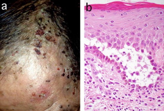
LON late onset neutropenia from rituximab...

EARLY RECOGNITION OF RITUXIMAB-INDUCED LATE ONSET NEUTROPENIA IS ESSENTIAL


DII small banner

By Warren R. Heymann, MD, FAAD
April 13, 2022
Vol. 4, No. 15

Rituximab is an anti-CD20 chimeric antibody directed against a surface transmembrane protein marker (CD20) expressed on B-cells during differentiation from pre B-cell until the plasma cell stage, involved in the maturation and activation of B cells. Approved in 1998, rituximab's off-label use has been expanding beyond its other FDA-approved indications (CD-20 positive B-cell non-Hodgkin lymphoma, chronic lymphocytic leukemia, rheumatoid arthritis, microscopic polyangiitis, and granulomatosis with polyangiitis). The list of off-label uses of rituximab continues to increase exponentially; from a dermatological perspective, its use in bullous pemphigoid, dermatomyositis, systemic lupus erythematosus, and refractory graft-versus-host disease are just a few examples where the drug may be impactful. (1)

Rituximab's well-recognized adverse events include infusion reactions, lymphopenia, hepatitis B virus reactivation, and progressive multifocal leukoencephalopathy. (2) Early-onset neutropenia is extremely rare, with only a handful of cases reported to date. (3) Rituximab-induced late-onset neutropenia (R-LON), although underrecognized, has been increasingly reported. According to the National Cancer Institute common toxicity criteria, R-LON is defined as unexplained grade 3 or more neutropenia (<1.0×10³/μL) occurring at least 3–4 weeks after the rituximab infusion. (4) Other authors define R-LON as (<1.5×10³/μL) after at least 4 weeks — the end point is not well-defined, being reported as late as 2 years following treatment.

Increasingly, extended courses of rituximab are being utilized to keep patients in remission. Rashid et al have demonstrated how maintenance infusions have prevented relapses of pemphigus. (5) According to Hosp et al, "LON is not infrequent. Its incidence is estimated at approximately 6.0% but might vary within a broad range (1.3% –29.9%). The risk of LON possibly depends on the underlying disease, and is supposed to be highest in coexisting lupus erythematosus. Comedication with disease-modifying antirheumatic drugs does not seem to influence its incidence." (6) Zonozi et al identified 107 episodes of LON in 71 patients. The cumulative incidence at 1 year was 6.6% (the incidence rate in the first year was higher compared to thereafter). Systemic lupus erythematosus and combination therapy with cyclophosphamide were each independently associated with an increased risk of LON. (7)

Illustration for DWII on early recognition of Rituximab-induced late onset neutropeniaJAAD 2005; 52: 839-845.

Is R-LON clinically significant? In their review, Hosp et al observed that although patients are frequently asymptomatic, severe infectious complications, including fatalities, have occurred. Administration of filgastrim (G-CSF) has been demonstrated to shorten time to recovery but did not affect overall outcomes. (6) Zonozi et al reported that fever and sepsis complicated 31.3% and 8.5% of episodes, respectively. Most patients (69%) were treated with filgrastim. Rituximab rechallenge occurred in 87% of patients, of whom 21% developed recurrent LON. (7) Boch et al identified LON in 5 of 117 rituximab-treated pemphigus patients compared to none of 15 patients who did not receive the drug. The R-LON in their patients was short-lived; none required specific therapy (antibiotics, filgastrim) or hospitalization. (8)

The pathomechanism(s) of R-LON remain to be determined. Hypotheses include the production of antineutrophil antibodies, expansion of large granular lymphocyte cells and aberrant B-cell reconstitution, and the association of an FcgRIIIa polymorphism. (2) Immunodysregulation during B-cell recovery coinciding with excess levels of B-cell activating factor (BAFF) and a proliferation-inducing ligand (APRIL) may be at play. (6) The presumption is that B-cell depletion leads to alterations in hematopoietic growth factors that promote lymphopoiesis and downregulate granulopoiesis. (7)

More will be learned about R-LON with increasing experience and study. Dermatologists must be aware of this adverse reaction of rituximab and monitor patients accordingly. While most patients will be fine, that is not a guarantee, and vigilance in recognizing severe infection is mandatory. As the sage Yogi Berra eloquently stated: "It gets late early out there."

Point to Remember: Late-onset neutropenia has been increasingly recognized in patients receiving rituximab. Although usually short-lived and without clinical consequence, severe infections and fatalities have been reported. Dermatologists need to be cognizant of this adverse reaction and monitor patients carefully during their course of rituximab therapy.

Our expert's viewpoint

Robert A. Somer, MD
Professor of Medicine, Cooper Medical School of Rowan University
Head, Division of Hematology and Medical Oncology; Executive Director, Clinical Trials Program
MD Anderson Cancer Center at Cooper

Dermatologists must be aware of the risks of rituximab, as use of this agent increases, not only for the treatment of pemphigus vulgaris, but other dermatologic conditions as mentioned above. As use of rituximab expands to treat multiple autoimmune disorders, sub-specialists will need to remain cognizant of the risks and benefits of this agent and monitor CBCs not only during the course of treatment but also thereafter. LON is much more common than we initially thought, and often found incidentally. Patients with pemphigus vulgaris may be at higher risk of cellulitis compared to other indications, if they experience rituximab associated LON. Luckily, most cases reverse and are unlikely to require therapy. If severe infections or sepsis do exist, treatment with G-CSF may be necessary. As more dermatologists get comfortable prescribing this agent for infusion, they must remain vigilant in monitoring. More investigation is certainly needed to accurately define the mechanism of LON which can then lead to prevention and treatment strategies as rituximab use continues to increase.

  1. Hanif N, Anwer F. Rituximab. 2020 Nov 3. In: StatPearls [Internet]. Treasure Island (FL): StatPearls Publishing; 2020 Jan–.

  2. Sahu KK, Petrou N, Cohn Z, Khanna S. Rituximab-induced late-onset neutropenia. BMJ Case Rep. 2019 Dec 29;12(12):e233569.

  3. Shah S, Kavadichanda CG, Belani P, Ganesh RN, Negi VS. Early onset neutropenia and thrombocytopenia following rituximab in lupus nephritis. Int J Rheum Dis. 2019 May;22(5):946-950.

  4. Sahu KK, Petrou N, Cohn Z, Khanna S. Rituximab-induced late-onset neutropenia. BMJ Case Rep. 2019 Dec 29;12(12):e233569.

  5. Rashid H, Lamberts A, van Maanen D, Bolling MC, Diercks GFH, Pas HH, Jonkman MF, Horváth B. The effectiveness of rituximab in pemphigus and the benefit of additional maintenance infusions: Daily practice data from a retrospective study. J Am Acad Dermatol. 2020 Nov;83(5):1503-1505.

  6. Hosp C, Goebeler M, Benoit S, Stoevesandt J. Rituximab-mediated late onset neutropenia in autoimmune blistering diseases: negligible or under-estimated threat? J Dtsch Dermatol Ges. 2020 Oct;18(10):1162-1164.

  7. Zonozi R, Wallace ZS, Laliberte K, Huizenga NR, Rosenthal JM, Rhee EP, Cortazar FB, Niles JL. Incidence, Clinical Features, and Outcomes of Late-onset Neutropenia from Rituximab for Autoimmune Disease. Arthritis Rheumatol. 2020 Sep 6. doi: 10.1002/art.41501. Epub ahead of print. PMID: 32892495.

  8. Boch K, Zillikens D, Langan EA, Schmidt E, Ludwig RJ. Low prevalence of late-onset neutropenia after rituximab treatment in patients with pemphigus. J Am Acad Dermatol. 2020 Dec;83(6):1824-1825.


All content found on Dermatology World Insights and Inquiries, including: text, images, video, audio, or other formats, were created for informational purposes only. The content represents the opinions of the authors and should not be interpreted as the official AAD position on any topic addressed. It is not intended to be a substitute for professional medical advice, diagnosis, or treatment.