Dermatología en Costa Rica

Wednesday, June 22, 2022

Dieta keto beneficios

La dieta cetogénica muy baja en calorías se perfila como una opción ventajosa para el abordaje del síndrome de ovario poliquístico

MADRID, ESP. Especialistas analizaron el papel de la dieta cetogénica muy baja en calorías(very-low-calorie ketogenic diet) en tres comorbilidades que tienen una incidencia más alta entre los pacientes con sobrepeso y obesidadsíndrome de ovario poliquísticohígado graso y diabetes de tipo 2, durante el Simposio Científico Internacional New frontiers in scientific research, celebrado recientemente en Barcelona, con el objetivo de hacer un análisis y puesta al día de las últimas evidencias sobre los beneficios de esta pauta dietética.[1]

Disfunción del tejido adiposo, obesidad y síndrome de ovario poliquístico

Dra. Alessandra Gambineri/Fuente: Pronokal Group

La Dra. Alessandra Gambineri, profesora asociada del Departamento de Medicina y Cirugía (DIMEC) de la Università di Bologna, en Bolonia, Italia, abordó el nexo entre la obesidad y el síndrome de ovario poliquístico, "una enfermedad crónica que afecta a cerca de 10% de las mujeres en edad fértil y que presenta fenotipos muy diversos con características distintas".

"La fisiopatología de este síndrome se caracteriza por la interacción de tres factores: exceso de andrógenos, disfunción de los tejidos adiposos y resistencia a la insulina, que interactúan entre sí y que se expresan de distinta manera en los diferentes fenotipos", puntualizó.

La especialista señaló que la disfunción del tejido adiposo es muy importante en esta patología, ya que está asociada a factores que se secretan, como ácidos grasos libres, citocinas proinflamatorias, algunas adipocinas que fomentan la resistencia a la insulina, glucocorticoesteroides, andrógenos y estrés oxidativo.

"Asimismo, el estrés oxidativo que caracteriza este síndrome se expresa todavía más cuando hay obesidad, que también produce una hipotoxicidad en el ovario que agrava la función ovulatoria. En este contexto, la dieta cetogénica muy baja en calorías puede ser útil en varios sentidos: reducción de peso, favoreciendo la pérdida de masa grasa, principalmente la visceral/abdominal. disminución de la lipotoxicidad y mejora del estado inflamatorio, la hiperinsulinemia y la resistencia a la insulina".

Esta fue la línea seguida para la realización del estudio* cuyos resultados expuso la Dra. Gambineri y que tuvo como objetivo analizar los efectos de la dieta cetogénica muy baja en calorías sobre las manifestaciones del síndrome de ovario poliquístico en el fenotipo de obesidad.

"La finalidad fue comparar los efectos de una dieta cetogénica muy baja en calorías y la dieta estándar baja en calorías (hipocalórica) como grupo control sobre peso corporal, resistencia a la insulina, ciclo menstrual, ovulación, morfología ovárica e hiperandrogenismo en una población de 30 mujeres con obesidad, síndrome de ovario poliquístico y resistencia a la insulina".

Las participantes del estudio tenían un diagnóstico de síndrome de ovario poliquístico según los criterios de National Institutes of Health (NIH) y edades entre 18 y 45 años; se les aleatorizó en dos grupos (15:15): experimental (dieta cetogénica muy baja en calorías) y control (dieta hipocalórica). "Las mujeres asignadas al grupo experimental siguieron la etapa cetogénica durante ocho semanas y posteriormente pasaron a la segunda fase con dieta hipocalórica durante ocho semanas más, mientras que las del grupo control (dieta hipocalórica) siguieron la dieta hipocalórica durante las 16 semanas".

Los resultados primarios fueron cambios en el peso y en la composición corporal, concretamente la masa grasa y la masa magra, medidas mediante bioimpedancia. "Como resultados secundarios se consideraron los cambios observados en distintos aspectos: distribución de la grasa abdominal, parámetros metabólicos, ovulación, morfología del ovario, hirsutismo, hiperandrogenismo, bienestar psicológico y distrés psicológico. También se analizó cómo se había reducido el estroma del ovario, la zona donde se sintetizan los andrógenos".

Los autores del estudio comprobaron que si bien el índice de masa corporal se redujo en ambos grupos, esta reducción fue mayor en el que siguió la dieta cetogénica muy baja en calorías. Se observó una pérdida de peso importante en ambos grupos, 12,4 kg frente a 4,7 kg. También se observaron diferencias significativas en otros parámetros: circunferencia de cintura (-8,1% en el experimental frente a -2,2% en el control), masa grasa (-15,1% frente a -8,5%) y testosterona libre (-30,3% frente a +10,6%).

En cuanto a la insulina, solo se produjo una reducción en el grupo experimental.

"Un dato muy importante es que respecto al hiperandrogenismo, sobre todo en lo que se refiere a la testosterona libre, se produjo una reducción significativa solo en el grupo de la dieta cetogénica muy baja en calorías y esta reducción se hizo especialmente patente en la primera parte del estudio, coincidiendo con el periodo cetogénico. La razón de este efecto está en el importante aumento de la concentración de las globulinas fijadoras de hormonas sexuales, las SHB6, que se unen a la testosterona presente en la sangre femenina, produciendo reducción de la testosterona libre, un efecto muy importante teniendo en cuenta que este síndrome es un trastorno androgénico y también que los tratamientos actuales para el síndrome de ovario poliquístico no consiguen reducir tanto la testosterona libre como lo hace este método dietético", señaló la Dra. Gambineri.

Para la especialista, entre todos estos efectos positivos en estas pacientes tal vez el más importante sea la gran mejora que se produce en la ovulación. "Al comienzo del estudio solo 38,5% de las participantes del grupo experimental y 14,3% de las del grupo de control tenían ciclos ovulatorios. Tras la intervención 84,6% consiguió ovular frente a 35,7% que logró este objetivo en el otro grupo".

La Dra. Gambineri sugiere que este método es "válido para reducir la masa grasa y mejorar rápidamente el hiperandrogenismo y la disfunción ovulatoria en mujeres con obesidad y síndrome de ovario poliquístico".

Mejora del perfil glucémico… ¿y reversión de la diabetes de tipo 2?

La Dra. Daniela Sofrà, endocrinóloga especialista en diabetología de la Clinique de La Source, en Lausanna, Suiza, repasó las evidencias de las que se dispone actualmente sobre el papel de la dieta cetogénica muy baja en calorías en el abordaje de la diabetes de tipo 2.

"Es el momento de repensar el tratamiento de la diabetes y centrar los esfuerzos en el manejo de la obesidad como factor asociado. Una de las hipótesis en las que se trabaja en este sentido es la del ciclo gemelo, que postula que la diabetes de tipo 2 es el resultado del exceso de grasa en el hígado, lo que a su vez se asocia a la resistencia a la insulina con disfunción del páncreas".

La Dra. Sofrà agregó que hay un estudio que documenta por primera vez la reversibilidad de la morfología del páncreas diabético tras una restricción calórica con la dieta cetogénica muy baja en calorías.[2] "La razón de este efecto es la utilización de la grasa visceral e intrahepática, que puede llevar a la remisión de la manifestación clínica de la diabetes de tipo 2, entendiendo por tal la definición que hace la American Diabetes Association (ADA): hemoglobina glucosilada < 6,5% sin terapia farmacológica".

Concretamente, los resultados de esta investigación mostraron que tras seguir la dieta cetogénica muy baja en calorías y conseguir con ella una pérdida de 15% del peso (media de pérdida ponderal de las participantes), los niveles de la glucosa hepática recuperaron niveles normales en siete días y que la función de las células beta volvió a ser casi normal en ocho semanas.

"Estudios posteriores han demostrado la durabilidad de la remisión de la diabetes de tipo 2 gracias a la reactivación de la función secretora de la insulina de las células beta que se habían desdiferenciado frente al exceso crónico de nutrientes. Concretamente, seis de cada diez pacientes mantenían la hemoglobina glucosilada < 6% después de seis meses sin necesidad de terapia farmacológica", agregó.

Asimismo, la especialista destacó que la probabilidad de lograr la remisión está determinada principalmente por la duración de la enfermedad: "Los años de diabetes son uno de los principales predictores de la respuesta que el paciente va a tener con esta intervención dietética. Estudios han demostrado que la remisión es posible en pacientes con diabetes de menos de seis años, aunque hay otros trabajos que apuntan que puede conseguirse hasta con diez años de duración".

Con base en estos datos, la Dra. Sofrà hizo hincapié en los efectos pleiotrópicos de la dieta cetogénica muy baja en calorías sobre el control de la glucemia, favoreciendo la posible remisión de la diabetes o la reducción de fármacos, así como la reducción del índice HOMA-IR (resistencia a la insulina) y de la circunferencia abdominal en personas con diabetes de tipo 2.

Terapia potencial para el hígado graso no alcohólico

La tercera comorbilidad de la obesidad que puede beneficiarse de la dieta cetogénica muy baja en calorías es la esteatosis hepática o enfermedad del hígado graso no alcohólico, expuso el Dr. Hardy Walle, especialista en Medicina Interna y director y fundador del centro Bodymed AG, en Kirkel, Alemania y uno de los autores de esta investigación.

"Investigaciones recientes muestran que la grasa ectópica y en particular la enfermedad del hígado graso no alcohólico podrían considerarse una causa —o al menos una de ellas— de la mayoría de las enfermedades que afectan a la población como consecuencia del sobrepeso y la obesidad, hasta el punto de que algunos autores han afirmado que sin hígado grado no hay diabetes de tipo 2", destacó.

El Dr. Walle señaló que entre 30% y 40% de la población adulta tiene enfermedad del hígado graso no alcohólico, un porcentaje que aumenta considerablemente en personas con obesidad, llegando a 70% de prevalencia y aumentando, en el caso de la diabetes de tipo 2, a casi 90%. "Incluso el normopeso no excluye de padecer hígado graso; de hecho, alrededor de 15% de las personas con enfermedad del hígado graso no alcohólico no tiene sobrepeso".

En un escenario en que no existen fármacos aprobados para el tratamiento del hígado graso (el abordaje estándar actual se centra en las intervenciones en el estilo de vida), una dieta hipocalórica a corto plazo (o ayuno hepático) se considera un método eficaz para el manejo de esta patología, como demostró un estudio de la Universität des Saarlandes, en Saarland, Alemania, comentado por el experto para ilustrar esta afirmación.

"Los participantes (60 pacientes con esteatosis hepática) siguieron una dieta hipocalórica (de menos de 1.000 kcal/día) durante 14 días con una fórmula rica en proteínas y fibras especialmente desarrollada para el tratamiento de la enfermedad del hígado graso no alcohólico. Después se realizó elastografía (fibroscan) con medición del parámetro de atenuación controlada para cuantificar la enfermedad del hígado graso.[3] Los resultados demostraron no solo una mejora significativa de los parámetros de la enfermedad del hígado graso no alcohólico, sino que también mejoraron de forma notable todos los parámetros metabólicos relevantes (lípidos séricos, enzimas hepáticas)", explicó el Dr. Walle.

"Estas evidencias nos llevan a afirmar que el concepto de ayuno hepático (mediante una dieta hipocalórica) marca un punto de referencia para una futura terapia de abordaje de la enfermedad del hígado graso no alcohólico, concluyó el especialista.

*Estudio llevado a cabo con la colaboración de Pronokal Group (Nestlé Health Science).Los doctores Gambineri, Sofrà y Walle han declarado no tener ningún conflicto de interés económico pertinente.

Siga a Carla Nieto de Medscape en español en Twitter @carlanmartinez.

Para más contenido siga a Medscape en FacebookTwitterInstagram y YouTube.


Sent from my iPhone

Benjamin Hidalgo-Matlock
Skin Care Physicians of Costa Rica

Clinica Victoria en San Pedro: 4000-1054
Momentum Escazu: 2101-9574

Please excuse the shortness of this message, as it has been sent from
a mobile device.

Tuesday, June 21, 2022

Nuevos Anticuerpos para LES y Esclerodermia.

New Blood Tests Approved for Lupus, Systemic Sclerosis Diagnoses

Carolyn Crist

June 17, 2022

The Food and Drug Administration has approved a new diagnostic blood test to detect monospecific Ribosomal P antibodies to support a diagnosis of systemic lupus erythematosus (SLE), particularly for people who are antinuclear antibody-negative, according to a recent announcement from the test developer, Thermo Fisher Scientific Inc.

The company also said the agency approved a separate blood test for RNA Polymerase III antibodies to support a diagnosis of systemic sclerosis (SSc), or scleroderma. RNA Polymerase III antibodies are a criteria marker for SSc with both diagnostic and prognostic value.

The company noted that a subset of patients with SLE present with ribosomal P antibodies, and the new test, called EliA Rib-P, aims to improve on the "unreliable method" of detecting these antibodies via indirect immunofluorescence, "which if used solely, could result in delayed diagnosis and treatment."

The company also noted that up to 70% of patients who test positive for RNA Polymerase III antibodies with the SSc test, called EliA RNA Pol III, have no other SSc-associated antibodies. The test also "completes a criteria-based EliA SSc panel and is the first fully automated RNA Polymerase test available" in the United States.

"The availability of a strong CTD test menu on a fully automated instrument could improve the efficiency and productivity of diagnostic laboratories," Dr Henry Homburger, Professor Emeritus of Laboratory Medicine at Mayo Clinic College of Medicine, and laboratory director, Thermo Fisher Phadia Immunology Reference Laboratory, said in the announcement. "The new EliA RNA Pol III and EliA Rib-P tests have been designed to improve the differentiation of SSc and SLE from other connective tissue diseases. Targeting existing diagnostic care gaps can potentially lead to earlier and more accurate diagnosis and ultimately improve clinical outcomes for patients."

Sources:

Lupus Foundation of America: "New Blood Test Approved for Lupus Diagnosis."

Thermo Fisher Scientific: "FDA Clears Thermo Scientific EliA RNA Pol III and EliA Rib-P Tests for Use in U.S."

Dermatitis atopica junto con diagnóstico de alergia alimentaria, No inducida por los alimentos.

Atopic dermatitis common among children with food allergies but rarely triggered by food

By Richard Gawel

ADD TOPIC TO EMAIL ALERTS

Receive an email when new articles are posted on

Please provide your email address to receive an email when new articles are posted on .

We were unable to process your request. Please try again later. If you continue to have this issue please contact customerservice@slackinc.com.

Although food allergy appeared common among children with atopic dermatitis, only 3% had food-triggered atopic dermatitis, according to a study published in The Journal of Allergy and Clinical Immunology: In Practice.

Because of the risks of unnecessarily eliminating foods, including nutritional deficiencies and loss of tolerance, detailed food allergy histories should be obtained from patients with atopic dermatitis, including testing and oral food challenges if indicated, Jennifer Chen Li, MD, an allergy/immunology fellow with the Massachusetts General Hospital division of rheumatology, allergy and immunology, and colleagues wrote.

Food-triggered atopic dermatitis occurred among 2% of patients with mild AD and food allergy, 6% of patients with moderate AD and food allergy and 4% of patients with severe AD and food allergy.
Data were derived from Li JC, et al. J Allergy Clin Immunol Pract. 2022;doi:10.1016/j.jaip.2022.05.028.

To distinguish the prevalence of food-triggered AD from IgE-mediated food allergy, the researchers conducted a retrospective chart review of 372 children with AD who were referred to allergy and/or dermatology specialists at a tertiary care referral center with one or more follow-up visits.

The children (63% boys; 76% white; 92% non-Hispanic) had a median age at first specialist visit of 1.1 years (range, 0 to 16 years), with 29% diagnosed with moderate AD and 18% diagnosed with severe AD.

Most patients with AD (55%) had IgE-mediated food allergy, including 60% of those with mild AD, 45% of those with moderate AD and 57% of those with severe cases of AD. About two-thirds (67%) of patients with food allergy were boys, and a greater proportion of patients with vs. without IgE-mediated food allergy were aged younger than 1 year at the initial visit (48% vs. 26%; P < .001).

The most common allergens associated with immediate-type food allergy included peanut (44%) and egg (43%). Also, 65% of patients with IgE-mediated food allergy had skin prick test or IgE levels with positive predictive values greater than 95%.

Food-triggered AD (FTAD) — defined by a physician-noted sustained improvement in AD after removal of a food — appeared uncommon, occurring in only 3% of the total cohort and 2% of patients with mild AD, 6% of those with moderate AD and 4% with severe AD.

Additionally, 4% of patients with an IgE-mediated food allergy to at least one food had FTAD for another food.

The most common allergens for FTAD were egg (62%) and peanut (31%). OFC was used to confirm the diagnosis of approximately 30% of patients with FTAD.

Among the 97 patients who only were referred for AD and not food allergy, 29% had IgE-mediated food allergy, 13% had mild AD, 28% had moderate AD and 41% had severe AD, while 5% developed FTAD.

A very small number of patients may experience better AD outcomes with food elimination diets, the researchers continued, but risks include nutritional deficiencies and loss of tolerance.

Providers should then carefully consider prescribing food elimination diets for AD when there are no immediate food allergy symptoms, with clinical decision-making focused on symptoms of IgE-mediated food allergy in most cases.

Even among patients who only were referred for AD, the researchers concluded, food allergy was common, although FTAD was rare. Noting that their study was retrospective, the researchers also said that prospective studies are necessary to further characterize the connection between food allergy and AD.

Read more about

ADD TOPIC TO EMAIL ALERTS

Receive an email when new articles are posted on

Please provide your email address to receive an email when new articles are posted on .

We were unable to process your request. Please try again later. If you continue to have this issue please contact customerservice@slackinc.com.

Monday, June 13, 2022

Síntomas de Rosacea Ocular

Published in Dermatology

Journal Scan / Research · June 12, 2022

Clinical Clues to Identify Patients With Ocular Rosacea

International Journal of Dermatology


TAKE-HOME MESSAGE

Abstract


BACKGROUND AND OBJECTIVES

Ocular rosacea is a special manifestation of rosacea with unknown etiology. Eye involvement in rosacea patients is surprisingly common; however, it is often underdiagnosed, resulting in inappropriate treatment. We aimed to provide an updated epidemiologic perspective on ocular rosacea in Germany to improve patient care.

PATIENTS AND METHODS

Data of 777 rosacea patients were assessed using a detailed online questionnaire regarding ocular and skin symptoms, previous dermatological and ophthalmological consults, presence of type 1 hypersensitivities, and Demodex testing. All data were statistically analyzed.

RESULTS

Most patients reported ocular symptoms (399/777, 51.4%), including red eyes (179/399, 44.9%), itching (187/399, 46.9%), sty or chalazion (309/399, 77.4%), and dryness (108/399, 27.1%). Ocular rosacea was confirmed in 149/309 cases who consulted an ophthalmologist (45.3%). A total of 159/399 (39.8%) had no pre-existing allergies. Eye involvement was significantly associated with the presence of skin symptoms (P < 0.05), impacting patients' general well-being and overall treatment satisfaction. About half of Demodex-positive patients (21/45, 46.7%) showed ocular symptoms.

CONCLUSIONS

Eye involvement in rosacea patients was common, often presenting with unspecific symptoms.


International Journal of Dermatology
Clinical clues to identify patients with ocular rosacea - a Germany-wide epidemiologic analysis
Int. J. Dermatol 2022 May 17;[EPub Ahead of Print], S Zierl, JA Hildebrand, A Guertler, C Dietrich, BM Clanner-Engelshofen, LE French, M Reinholz 

Sent from my iPhone

Benjamin Hidalgo-Matlock
Skin Care Physicians of Costa Rica

Clinica Victoria en San Pedro: 4000-1054
Momentum Escazu: 2101-9574

Please excuse the shortness of this message, as it has been sent from
a mobile device.

Sunday, June 12, 2022

Acne y Enfermedad Inflamatoria Intestinal

Published in Dermatology

Journal Scan / Research · June 11, 2022

Genetic Liability to Acne Is Associated With Increased Risk of Inflammatory Bowel Disease

Journal of the American Academy of Dermatology

TAKE-HOME MESSAGE

Abstract

Isotretinoin, an effective mediation prescribed for severe acne, has been suspected to trigger inflammatory bowel disease (IBD). However, with more recent evidence that supports null association between isotretinoin exposure and IBD risk, there is a growing speculation as to whether IBD may in fact be part of the consequences of acne itself, rather than the side-effect of isotretinoin treatment. Only 1 population-based study has previously touched on this hypothesis, indicating a direct effect of acne on IBD. As the causal association is difficult to establish depending solely on classical observational designs, we performed a 2-sample Mendelian randomization (MR) study to test the hypothesis that acne would increase the risk of IBD. MR leverages randomly allocated genetic variants as instruments (Fig 1), which could infer the causal effect of an exposure on an outcome by minimizing confounding and reverse causality.

Journal of the American Academy of Dermatology
Genetic liability to acne is associated with increased risk of inflammatory bowel disease: A Mendelian randomization study
J Am Acad Dermatol 2022 May 02;[EPub Ahead of Print], J Zhu, D Zhou, J Wei, Y Li 


Sent from my iPhone

Benjamin Hidalgo-Matlock
Skin Care Physicians of Costa Rica

Clinica Victoria en San Pedro: 4000-1054
Momentum Escazu: 2101-9574

Please excuse the shortness of this message, as it has been sent from
a mobile device.

Thursday, June 02, 2022

Monkey Pox

THE GLOBALIZATION OF MONKEYPOX


DII small banner

By Warren R. Heymann, MD, FAAD
May 25, 2022
Vol. 4, No. 21

Disclaimers: This commentary was written on May 22 for a publication date of May 25, 2022. The issues related to the increasing number of reported monkeypox cases are changing rapidly. The content included in this commentary may be outdated by the time of publication. The reader is encouraged to stay abreast of developments via the CDC as well as local government and institutional health care authorities. 

Here we go again. I have not thought much about monkeypox (MP) since 2003 when a documented case appeared in a 3-year-old girl from central Wisconsin. Investigations implicated a shipment of 800 small animals from Ghana to Texas as the probable means of entry of the MP virus into the United States. According to Sale et al: "A sick Gambian giant-pouched rat (Cricetomys gambianus) from this shipment was subsequently sold to an Illinois animal vendor where it was kept in close proximity to prairie dogs (a native North American rodent [Cynomys species]). Prairie dogs that became infected were sold to a second animal distributor, and eventually to two pet shops and at a pet swap meet in northern Wisconsin. Additional animals from the Texas shipment, including dormice and rope squirrels, tested positive for monkeypox virus. As of July 2003, 72 cases of human monkeypox were under investigation in Illinois, Indiana, Kansas, Missouri, Ohio, and Wisconsin with 37 cases confirmed through laboratory analysis." (1)

As of today, a case has been identified in Massachusetts along with two in Canada. (2) Kozlov states: "More than 120 confirmed or suspected cases of monkeypox, a rare viral disease seldom detected outside of Africa, have been reported in at least 11 non-African countries in the past week. The emergence of the virus in separate populations across the world where it doesn't usually appear has alarmed scientists — and sent them racing for answers." (3) 

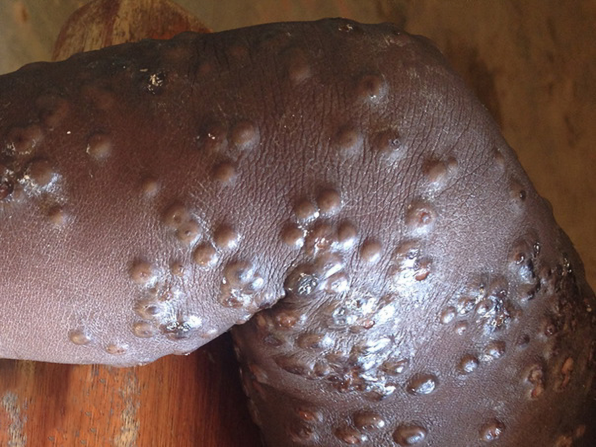
Image for DWII on monkeypox
Image from DermNetNZ.

(The image above shows monkeypox on a darker skin tone. NPR recently published an image of monkeypox on white skin, which can be seen in a recent article.)

There is no need for panic — MP is not analogous to COVID-19. A pandemic from MP is not in the offing. Regardless, dermatologists must be familiar with all aspects of MP in order to identify possible cases which may assist in limiting contagion of this potentially fatal disease. This scenario reminds me of the anthrax scare following 9/11. I did not see a single case but had to evaluate dozens of patients who were convinced they were infected with Bacillus anthracis. (Most of those patients had impetigo or insect bites). Regardless, dermatologists needed to be prepared for the possibility of encountering the disease. The same holds true for MP. This commentary will take a question and answer format, followed by an expert opinion by Misha Rosenbach, MD, FAAD.

What is MP?

MP is a rare, potentially life-threatening zoonotic infection that occurs predominantly in West and Central Africa. It is caused by the monkeypox virus, a double-stranded DNA orthopoxvirus similar to the variola virus (the causative agent of smallpox) and vaccinia virus (the live virus component of orthopoxvirus vaccines). (4,5)

When was MP first recognized?

MP was first detected in 1958 in an outbreak of a vesicular disease in captive monkeys transported to Copenhagen, Denmark from Africa for research purposes. The term is actually a misnomer because the largest animal reservoirs of the MP virus are found in rodents, not monkeys. (5) The first human case of MP was reported in 1970, in a 9-year-old boy from Zaire (now known as the Democratic Republic of Congo), presenting with a smallpox-like disease from which MP virus was isolated. (5)

Where is MP usually found?

Ultimately, several thousand human cases of monkeypox have been confirmed in 15 different countries, with 11 of them in African countries. Monkeypox was imported to the United Kingdom, the U.S., Israel, and Singapore. (6) The increasing incidence of human MP cases in Central and West Africa is considered a function of diminished cross-protective immunity after smallpox vaccination was discontinued in the early 1980s following the eradication of smallpox. (5)

Are there variants of the MP virus? 

According to Moore and Zohra: "There are two distinct clades of the monkeypox virus. The West African clade has a more favorable prognosis with a case fatality rate below 1%. On the other hand, the Central Basin clade (Central African clade) is more lethal, with a case fatality rate of up to 11% in unvaccinated children." (7) Preliminary genetic data suggests that the MP virus is related to a viral strain predominantly found in western Africa. (3)

How is the MP virus transmitted?

The mode of transmission of the MP virus from animals to humans is unknown. Direct or indirect contact with live or dead animals is presumed to be responsible for most MP infections in humans. (5) Mahase states: "Transmission between people mostly occurs through large respiratory droplets, normally meaning prolonged contact face to face. But the virus can also spread through bodily fluids. The latest cases have mainly been among men who have sex with men. The UK Health Security Agency said that, although monkeypox has not previously been described as a sexually transmitted infection, it can be passed on by direct contact during sex. It can also be passed on through other close contact with a person who has monkeypox or contact with clothing or linens used by a person who has monkeypox." (8) Compared to the SARS-CoV-2 virus, which spreads by aerosol particles that stay aloft for minutes, the MP virus spreads through large droplets which fall to the ground within a few feet. (2)

What are the clinical manifestations of MP?

According to Petersen et al: "The incubation period has been estimated at 5 to 21 days, and duration of [signs and symptoms] at 2 to 5 weeks. The illness begins with nonspecific [manifestations] that include fever, chills, headaches, lethargy, asthenia, lymph node swellings, back pain, and myalgia (muscle ache) and begins with a fever before rashes appear. Within 1 to 5 days after the onset of fever, rashes of varying sizes appear, first on the face, then across the body, hands, and legs and feet. The rash undergoes several stages of evolution from macules, papules, vesicles (fluid-filled blisters), and pustules, followed by resolution over time with crusts and scabs, which drop off on recovery. Various stages of the rash may show at the same time. Areas of erythema and/or skin hyperpigmentation are often seen around discrete lesions. Detached scabs may be considerably smaller than the original lesion. Inflammation of the pharyngeal, conjunctival, and genital mucosae may also be seen." (5)

The main clinical differential diagnosis is smallpox. The lesions of MP appear monomorphic and pea-sized. The crop-like appearance of MP lesions, its less strong centrifugal spread, and presence of lymphadenopathy are important signs that differentiates MP from smallpox. (6)

What are potential complications of MP?

Complications may include secondary bacterial infections, sepsis, pneumonia, encephalitis, dehydration, cutaneous scarring, corneal scarring, post-inflammatory hyper or hypopigmentation, and death. (7)

What laboratory tests confirm the diagnosis of MP?

PCR (RT-PCR), serology (IgM and IgG), and electron microscopy have been utilized to confirm the diagnosis of MP. (6)

How are cases of MP treated?

As with most viral diseases, treatment is mostly supportive. Patients should be isolated with appropriate personal protective equipment (PPE). According to Moore and Zohra, "For severe cases, investigational use can be considered for compounds with demonstrated benefit against orthopoxviruses in animal studies and severe vaccinia vaccine complications. The oral DNA polymerase inhibitor brincidofovir, oral intracellular viral release inhibitor tecovirimat, and intravenous vaccinia immune globulin have unknown efficacy against the monkeypox virus."

People exposed to the virus should be monitored for 21 days. There is no need for isolation unless they become symptomatic. In some cases, post-exposure vaccination with modified vaccinia, Ankara vaccine (smallpox and monkeypox vaccine, live, non-replicating) is recommended. "Contact between broken skin or mucous membranes and an infected patient's body fluids, respiratory droplets, or scabs is considered a 'high risk' exposure that warrants post-exposure vaccination as soon as possible. According to the CDC, vaccination within four days of exposure may prevent disease onset, and vaccination within 14 days may reduce disease severity." (7) Because human-to-human transmission appears to be limited, "ring" vaccination of close contacts is a more likely scenario than vaccination of the public at large. (2)

What should dermatologists be doing about the increase of MP cases?

According to the CDC (9):

  • If health care providers identify patients with a rash that looks like monkeypox, consider monkeypox, regardless of whether the patient has a travel history to central or west African countries.

  • Do not limit concerns to men who report having sex with other men. Those who have any sort of close personal contact with people with monkeypox could potentially also be at risk for the disease.

  • Some patients have had genital lesions and the rash may be hard to distinguish from syphilis, herpes simplex virus (HSV) infection, chancroid, varicella zoster, and other more common infections [such as molluscum contagiosum]. [WRH – perform appropriate tests to rule out these clinical entities]

  • Isolate any patients suspected of having monkeypox in a negative pressure room, and ensure staff understand the importance of wearing appropriate PPE and that they wear it each time they are near suspected cases.

  • Consult the state health department or CDC's monkeypox call center through the CDC Emergency Operations Center (770-488-7100) as soon as monkeypox is suspected. In the coming weeks we will be learning about the extent of MP infections, and the science propagating the outbreaks. We need to be honest about what we know and what we do not. 

Point to Remember: There have been an increasing number of cases of monkeypox beyond Africa. Dermatologists should familiarize themselves with the disease and establish an in-office protocol for suspected cases. 

Our expert's viewpoint

Misha Rosenbach, MD, FAAD
Associate Professor of Dermatology at the Hospital of the University of Pennsylvania

Thank you, as always, Warren, for rapidly distilling critical information for dermatologists, and thank you, too, for inviting me to comment. First — I am not an expert on monkeypox! I am, however, an 'expert' on reading, reviewing the literature, medical education, and communicating things to different audiences. Second — no one should panic about monkeypox. This is not COVID. However, lessons learned — and ignored — over the past 2 years are important to keep in mind as we frame any discussion around this new (to us, in the U.S.) pathogen.

As with COVID, clear messaging to ensure people are aware of what they need to know, in a timely fashion, is essential. Early in the COVID pandemic, physicians used social media to share information quickly, particularly dermatologists in Italy and across the EU, through use of WhatsApp, which led to an incredible atlas of the spectrum of cutaneous morphologies that were being observed early in the first wave of the pandemic. (10) Similarly, social media has allowed rapid alerts across the globe so that folks who may encounter monkeypox are thinking about the infection. Rapid diagnosis is essential, as recognizing the infection allows isolation and (potentially) treatment of cases; moreover, it helps public health officials identify contacts, evaluate their risk for exposure, monitor them for symptoms, and (potentially) roll out ring vaccination. Notably, the mainstays of management are: informed frontline providers making a rapid diagnosis; trust and engagement with the health care system by cases and contacts; strong public health; adoption of vaccination by contacts. All of these have been eroded to some degree during the pandemic, whether due to fatigue, misinformation, or both. Some of that misinformation has come from physicians, which highlights the critical need for the field of medicine, including licensing boards, to actively combat medical misinformation (11, 12).

As an inpatient dermatologist who is frequently called to consult on new rashes in the emergency department, and founder of our urgent care access clinic in a major urban center and tertiary care academic referral center, I'm acutely aware of the need to disseminate accurate, up-to-date information to our colleagues and trainees. First, rapid recognition is essential. Dermatologic manifestations are common, and according to some papers, may be the initial signs of monkeypox infection. Warren reviewed this quite nicely up above — I will highlight that the CDC notes that manifestations start in the mouth and on the tongue, then progress outwards, generally evolving through stages together at each site. The morphologic evolution is reported to go from macules (1-2 days) to papules (1-2 days) to vesicles (1-2 days) to pustules (5-7 days) to eroded/crusted lesions. Patients frequently have a prodrome of fevers and other nonspecific symptoms, though lymphadenopathy is common (as opposed to smallpox). Lesions may be painful. The entire illness can last 2-4 weeks. Patients may also frequently have genital involvement. Note that many dermatologists are facile with the bedside diagnostic technique of Tzanck smears. We have employed these to distinguish herpes-family infections, such as HSV/VZV, from molluscum — another pox virus. Tzanck preparations performed on herpes infections will demonstrate multinucleated keratinocytes with nuclear molding and margination of chromatin. I have not seen a photograph of a Tzanck performed on monkeypox; however, Tzanck of molluscum can show oval-shaped bodies correlating with the Henderson-Patterson bodies seen on H&E stains of histologic sections (13). Importantly, use of high quality masks (N95 or equivalent), goggles, and contact precautions should protect the medical team during patient evaluation — and are another reminder of the importance of clear, accurate messaging around appropriate PPE.

A few important notes that were not covered above. First — it is true that some of the emerging groups of infection have been clustered in the MSM community. Monkeypox is not a gay disease, and it is essential to avoid stigmatization (14). Monkeypox is spread by contact or droplets — sexual contact is of course a risk factor. I would encourage readers to be thoughtful of potential issues around this, particularly in light of the various attacks and laws that are expanding against the LGBTQ community, and consider only careful messaging around this issue. Second — monkeypox is not a new disease. Our colleagues in Africa have been dealing with rolling outbreaks for years, and have extensive experience in managing the disease from a supportive care and public health standpoint. This is a good time to remind ourselves that humanity is interconnected — and to consider refocusing on neglected diseases, potential emerging pathogens, and issues of medical disparities. As the impact of the COVID pandemic still (and will) dwarf that of monkeypox, this is also a good time to remind readers about vaccine disparities, and the need to vaccinate the world against COVID. Many nations with experience battling monkeypox still lack sufficient vaccines against SARS-CoV-2, for instance. 

I would encourage every dermatologist to add monkeypox to the differential diagnosis when evaluating patients with new papulovesicular or vesiculopustular lesions. Particularly if they're occurring in crops, with a prodrome of fever and concurrent lymphadenopathy. Patients should be evaluated for exposure history, and infectious disease and public health colleagues engaged rapidly if the diagnosis is suspected or confirmed. In an ideal world, this new international outbreak should be rapidly contained. Doing so requires dermatologists to help make diagnoses quickly, and requires cases and their contacts to positively engage with the public health system. While it may seem premature to share information about monkeypox with only approximately 100 cases internationally, I hope we have all learned that early intervention and rapid containment is the best way to prevent pathogens from spiraling out of control. With the widespread emergence of monkeypox in multiple countries, if we react appropriately right now, at worst we might be accused of having overreacted — and that is the future I want. Accurate and timely information is essential — and I thank Warren, and the Dermatology World Insights & Inquiries team, for creating this so quickly.

  1. Sale TA, Melski JW, Stratman EJ. Monkeypox: an epidemiologic and clinical comparison of African and US disease. J Am Acad Dermatol. 2006 Sep;55(3):478-81. doi: 10.1016/j.jaad.2006.05.061. PMID: 16908354.

  2. Avril T. Monkeypox is almost nothing like COVID. Here's what to know, from two Phill scientists who've studied it.Philadelphia Inquirer, May 19, 2022. 

  3. Kozlov M. Monkeypox goes global: why scientists are on alert. Nature. 2022 May 20. doi: 10.1038/d41586-022-01421-8. Epub ahead of print. PMID: 35595996.

  4. Rao AK, Schulte J, Chen TH, Hughes CM, Davidson W, Neff JM, Markarian M, Delea KC, Wada S, Liddell A, Alexander S, Sunshine B, Huang P, Honza HT, Rey A, Monroe B, Doty J, Christensen B, Delaney L, Massey J, Waltenburg M, Schrodt CA, Kuhar D, Satheshkumar PS, Kondas A, Li Y, Wilkins K, Sage KM, Yu Y, Yu P, Feldpausch A, McQuiston J, Damon IK, McCollum AM; July 2021 Monkeypox Response Team. Monkeypox in a Traveler Returning from Nigeria - Dallas, Texas, July 2021. MMWR Morb Mortal Wkly Rep. 2022 Apr 8;71(14):509-516. doi: 10.15585/mmwr.mm7114a1. PMID: 35389974; PMCID: PMC8989376.

  5. Petersen E, Kantele A, Koopmans M, Asogun D, Yinka-Ogunleye A, Ihekweazu C, Zumla A. Human Monkeypox: Epidemiologic and Clinical Characteristics, Diagnosis, and Prevention. Infect Dis Clin North Am. 2019 Dec;33(4):1027-1043. doi: 10.1016/j.idc.2019.03.001. Epub 2019 Apr 11. PMID: 30981594.

  6. Alakunle E, Moens U, Nchinda G, Okeke MI. Monkeypox Virus in Nigeria: Infection Biology, Epidemiology, and Evolution. Viruses. 2020 Nov 5;12(11):1257. doi: 10.3390/v12111257. PMID: 33167496; PMCID: PMC7694534.

  7. Moore M, Zahra F. Monkeypox. 2022 Feb 28. In: StatPearls [Internet]. Treasure Island (FL): StatPearls Publishing; 2022 Jan–. PMID: 34662033.

  8. Mahase E. Monkeypox: What do we know about the outbreaks in Europe and North America? BMJ. 2022 May 20;377:o1274. doi: 10.1136/bmj.o1274. PMID: 35595274.

  9. https://www.cdc.gov/media/releases/2022/s0518-monkeypox-case.html

  10. Galván Casas C, Català A, Carretero Hernández G, et al. Classification of the cutaneous manifestations of COVID-19: a rapid prospective nationwide consensus study in Spain with 375 cases. Br J Dermatol. 2020;183(1):71-77. doi:10.1111/bjd.19163

  11. Baron RJ, Ejnes YD. Physicians Spreading Misinformation on Social Media — Do Right and Wrong Answers Still Exist in Medicine? [published online ahead of print, 2022 May 18]. N Engl J Med. 2022;10.1056/NEJMp2204813. doi:10.1056/NEJMp2204813

  12. Rubin R. When Physicians Spread Unscientific Information About COVID-19. JAMA. 2022;327(10):904-906. doi:10.1001/jama.2022.1083

  13. Wanat KA, Dominguez AR, Carter Z, Legua P, Bustamante B, Micheletti RG. Bedside diagnostics in dermatology: Viral, bacterial, and fungal infections. J Am Acad Dermatol. 2017;77(2):197-218. doi:10.1016/j.jaad.2016.06.034

  14. https://speakingofmedicine.plos.org/2022/05/19/monkeypox-is-not-a-gay-disease/ 


All content found on Dermatology World Insights and Inquiries, including: text, images, video, audio, or other formats, were created for informational purposes only. The content represents the opinions of the authors and should not be interpreted as the official AAD position on any topic addressed. It is not intended to be a substitute for professional medical advice, diagnosis, or treatment.

DW Insights and Inquiries archive

Explore hundreds of Dermatology World Insights and Inquiriesarticles by clinical area, specific condition, or medical journal source.


Sent from my iPhone

Benjamin Hidalgo-Matlock
Skin Care Physicians of Costa Rica

Clinica Victoria en San Pedro: 4000-1054
Momentum Escazu: 2101-9574

Please excuse the shortness of this message, as it has been sent from
a mobile device.