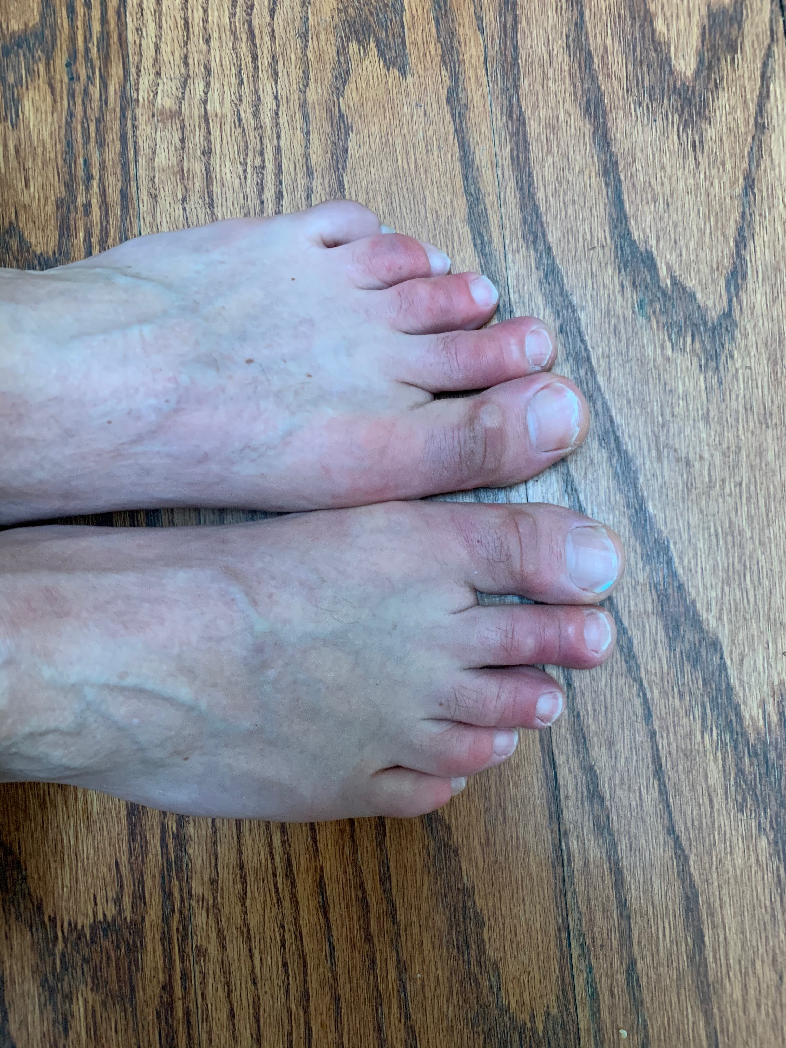
The profound dermatological manifestations of COVID-19: Part IV – Cutaneous features

By Warren R. Heymann, MD
April 22, 2020
Vol. 2, No. 16
Disclaimers: This commentary was written on April 17, 2020 for a publication date of April 22, 2020. The issues related to the COVID-19 pandemic are changing by the millisecond. The content included in this commentary may no longer be factual or relevant by the publication date. Part I of this series was referenced; Parts II and III were personal reflections; Part IV is referenced. The reader is encouraged to stay abreast of developments via the CDC and local government and institutional health care authorities. The AAD has instituted the valuable Coronavirus Resource Center and the COVID-19 Dermatology Registry to help understand the dermatologic manifestations of the COVID-19 virus.
It is hard to fathom that Part I of this series was published just a month ago. The rapid and complete transformation of our world health and economy is spellbinding, disorienting, heart wrenching, and devastating. Yet, despite the tribulations, the heroic determination to conquer the ravages of this novel coronavirus has been the impetus for the acquisition and dissemination of new knowledge.
In Part I, I quoted a study from China detailing 1099 patients with COVID-19 in which 2 patients (0.2%) had a "rash." (1) As I am writing this commentary there have been 3,541,368 cases and 153,379 deaths worldwide. As the global crisis has mushroomed, the international dermatological community has started to report relevant observations regarding the cutaneous manifestations of this affliction.
As viral information goes "viral," so do potential misconceptions. While trends are beginning to become apparent, the fact is that we are in the data-gathering phase of this epidemic, and dermatologists should not reach foregone conclusions based on scattered case reports in the medical literature or lay press. For example, in the article "What are Covid toes? Dermatologists, podiatrists share strange findings" (by Melissa Hohman, Today, April 17, 2020), it is stated that the pernio-like condition "seems more common in children and young people, but it's 'not exclusive' to them." Indeed, I had a curbside consult on a 78-year-old woman with severely purple toes and other symptoms raising the suspicion of COVID-19. We must be cognizant of confirmation bias — not every case of pernio need be associated with COVID-19 (the corollary being the apocryphal Freudian quote "sometimes a cigar is just a cigar"). Reports of cutaneous findings may not equate with association or causation. This week I had a teledermatology consult on a COVID-19 patient who clearly had chronic submammary intertrigo — I teased my resident: "Mike, you better write this up quickly as the first case of chronic intertrigo reported in COVID-19!"

The diagnosis of COVID-19 is based on clinical signs (fever, fatigue, dry cough, anorexia, dyspnea, rhinorrhea, ageusia, anosmia), on vital parameters (temperature, pulse oximetry saturation), and on radiological findings (X-ray, chest CT scan). Laboratory findings demonstrate lymphopenia and an elevated LDH. Viral isolation (utilizing PCR) by nasopharyngeal and oropharyngeal swabs confirms the diagnosis. Recalcati, a dermatologist in Italy, collected data on 88 COVID-19 patients — 18 patients (20.4%) developed cutaneous manifestations; 8 patients developed cutaneous involvement at the onset, 10 patients after hospitalization. Cutaneous manifestations were an erythematous rash (14 patients), widespread urticaria (3 patients), and chickenpox-like vesicles (1 patient). Lesions were mostly truncal. Pruritus was minimal or absent and lesions usually healed in a few days. There was no apparent correlation with disease severity. (2)
In their commentary about Recalcati's article, Su and Lee questioned whether the three patterns described (erythematous, urticarial, and varicelliform) are specific to the coronavirus. Additionally, they advocated further studies to determine the dynamic viral load and viremia at different points in the rash (prior, during, and after). (3) In a reply to Recalcati, Henry et al reported the case of a 27-year-old woman who presented with odynophagia, diffuse arthralgia, and pruritic disseminated erythematous plaques with facial and acral involvement, diagnosed as urticaria on dermatological consultation. Two days later she was febrile with chest pain, testing positive for COVID-19. (4) In a final reply, Estébanez et al reported the case of a 28-year-old woman who presented with cough, fatigue, and other symptoms, was found to be coronavirus-positive. Thirteen days after being tested she noticed pruritic lesions of the heels described as confluent "erythematous-yellowish" papules; 3 days later they appeared as pruritic, hardened, erythematous plaques. The clinical differential diagnosis included urticaria, urticarial vasculitis, idiopathic plantar hidradenitis, and neutrophilic dermatosis. No biopsy was obtained. (5)
Joob and Wiwanitkit presented a case from Thailand with a petechial eruption, initially considered to be Dengue fever. The patient subsequently developed respiratory problems which proved to be COVID-19. (6) In a reply to this report, Jiminez-Cauhe et al described an "erythemato-purpuric, millimetric, coalescing macules, located in flexural regions. The rash was mildly pruriginous and mainly located in the peri-axillary area." The authors believed this to be COVID-related but could not rule out the remote possibility that this was a drug rash due to administration of hydroxychloroquine and lopinavir/ritonavir at the time of admission. (7)
Manalo et al presented two cases of unilateral transient livedo reticularis (LR) in COVID-19 patients. The first patient was a 67-year-old man with right lower extremity LR that lasted "19 hours" and correlated with gross hematuria and generalized weakness. The other patient was a 47-year-old woman who noticed LR of her right leg after being outside for about 30 minutes. The rash lasted 20 minutes. Neither patient was critically ill. The authors hypothesized that the LR may have been due to microthromboses in these patients, possibly from a low-grade DIC. (8) Although I understand the postulate, it is hard to imagine that such transient eruptions are due to microemboli or thrombi — other factors must be at play. Antiphospholipid antibodies and coagulopathy resulting in lower extremity and hand digital ischemia accompanied by cerebral infarctions have been reported (9). Perhaps such coagulopathies could be contributing to LR or other acral pernio-like lesions.
Immunological factors presumably contribute to the pathogenesis of cutaneous lesions. According to Schett et al, COVID-19 "leads to fast activation of innate immune cells, especially in patients developing severe disease. Circulating neutrophil numbers are consistently higher in survivors of COVID-19 than in non-survivors, and the infection also induces lymphocytopenia that mostly affects the CD4+ T cell subset, including effector, memory, and regulatory T cells. Reflecting innate immune activation, levels of many pro-inflammatory effector cytokines, such as TNF, IL-1β, IL-6, IL-8, G-CSF, and GM-CSF, as well as chemokines, such as MCP1, IP10, and MIP1α, are elevated in patients with COVID-19, with higher levels in those who are critically ill. In addition, the levels of some T cell-derived cytokines, such as IL-17, are increased in the context of SARS-CoV-2 infection… In some patients with COVID-19, a cytokine storm develops that resembles secondary haemophagocytic lymphohistiocytosis, a hyperinflammatory state triggered by viral infections." (10)
As the COVID-19 pandemic continues (and hopefully abates in the coming weeks or months) the ongoing accrual of knowledge will give a clearer picture of how to interpret the panoply of cutaneous manifestations, both diagnostically and prognostically.
Point to Remember: We are in the initial phases of learning about the cutaneous manifestations of COVID-19. As expected, a viral type exanthem may be noted; additionally, acral pernio-like lesions, livedo reticularis, urticaria, petechial, and vesicular rashes have all been described. Further studies describing the clinical features of the infection, combined with basic research in its pathogenesis, will provide the foundation for eradication of this scourge.
Our Expert's Viewpoint
Misha Rosenbach, MD
Associate Professor of Dermatology
Hospital of the University of Pennsylvania
COVID-19, "COVID toes," and clinical manifestations
Disclosure: I am a member of the AAD Ad Hoc Task Force on COVID-19 and the Medical Dermatology Society's Ad Hoc Task Force, and involved in the AAD's COVID-19 registry referenced above (Director: Esther Freeman).
It is always an honor to be asked to comment on one of Warren's pieces, but it's also a challenge — particularly in this case, where he frequently took the words out of my mouth as I was reading his writing.
COVID-19 is a devastating disease with multiorgan manifestations. Science magazine titled a recent piece "How does coronavirus kill? Clinicians trace a ferocious rampage through the body, from brain to toes," and emerging reports have demonstrated that COVID-19 can impact almost every organ and system in the body, including the skin — and perhaps particularly, the toes.
However, Warren touched on another key point: not everything we see is COVID-19 related. With three million cases, if there were a striking, consistent rash, it would likely be well described by this point. Science has moved at blinding speed to answer the challenge of COVID-19, but that has also led to rapid acceptance or pre-print releases of articles which, in another era, may not get the attention that comes with having COVID-19 in the title. Social media has helped some misinformation proliferate (understatement of the year), but also served as a vehicle for frontline clinicians, including dermatologists, to rapidly share their observations and more rapidly adapt to managing patients with COVID-19. A brilliant colleague and friend wrote on March 30, 2020, "There's no COVID rash. We would know by now. If nearly a million cases of a disease occurs you are bound to have either misdiagnoses of other viruses, coincident drug rashes, or other random findings. Case series without pictures and path should not be published." He was, and is, likely correct: there is no universal COVID-19 rash.
However, there are clearly some skin manifestations in subsets of patients (which that same friend has also acknowledged!). The challenge we have as dermatologists is determining: 1) are any skin signs sensitive or specific enough to "count" as "presumed positive / past infection" with COVID-19; 2) are any skin signs clinically important in the acute management of patients; 3) do skin signs tell us about the pathophysiology of the disease; 4) can any skin signs sub-phenotype patients, leading to changes in management; 5) are there other plausible explanations for the skin findings.
Critically ill patients in ICUs have developed multi-organ thrombotic events, leading to questions of empiric anticoagulation in some patients with COVID-19. Another brilliant colleague, Dr. Joanna Harp (a "skin serious" dermatologist making a real difference on the front-lines), rounding daily in the ICU in heavily-hit Manhattan, observed skin changes ranging from livedo racemosa to retiform purpura, with intravascular thrombi on biopsy. (11) This may represent an important "skin sign of systemic disease" heralding intravascular, multiorgan clotting — and may suggest alternate, supplemental therapeutic options, as the authors speculate.
On the other end of the spectrum, "COVID toes," or acral, pernio-like lesions, seem to be more common in young patients, children and adolescents in particular, and portend a mild course, or develop after asymptomatic infection. As someone who cares for patients with sarcoidosis, I am used to "pernio" (lupus pernio) suggesting a different prognosis than other skin manifestations (in sarcoidosis, of course, lupus pernio implies more severe, chronic disease!). It will be essential to monitor this morphologic finding closely as antibody testing becomes available; if all patients with these pernio-like lesions are IgG positive and it becomes a presumptive sign of past infection, that has profound implications on testing. Given the inordinate, undeserved attention cast on hydroxychloroquine thus far, it pains me to raise this point, but as hydroxychloroquine is often employed as a therapeutic agent in treating pernio, one could speculate that if patients with COVID-19 are treated with hydroxychloroquine, even if they would have developed pernio post-infection, the anti-malarial therapy may suppress or abort the development of those characteristic lesions. Careful case definitions and evaluation of concomitant medications and alternate explanations for any observed pattern of skin findings remain essential in the evaluation of patients with COVID-19.
Additionally, it is important to consider alternate explanations for these cases of pernio. The weather in North America has been cooler this spring in parts of Canada and small regions of North America (though March overall was another record-warm month), due partially to an abnormal jet stream as a result of climate change. Idiopathic pernio is traditionally seen in cooler, damp climates — and it may be a coincidence that COVID-19 is devastating North America right as damp spring is occurring. Given the press surrounding "COVID-toes" and well-earned fear of COVID-19, patients with these sometimes subtle skin findings may be seeking more medical attention than they otherwise would for relatively innocuous lesions, leading to an uptick in diagnosis.
The urticarial, morbilliform, exanthematous eruptions described seem thus far both nonspecific and inconsistent enough to aid in either making a diagnosis or offering prognostic interpretations, but as with the rest, warrant monitoring. We as a field will need to closely evaluate the data, particularly that of the international registry effort being run by the AAD under the direction of Dr. Esther Freeman.
In the meantime, I hope you are all safe and healthy as can be, and advocating for more production of test kits, establishment of contact-tracing and isolation capabilities, and of course more personal protective equipment. It will take heroic efforts to overcome COVID-19, and we should be raising our voices to advocate for what is necessary to hold this disease in check until there are effective treatments and/or a vaccine.
Guan WJ, Ni ZY, Hu Y, Liang WH, et al. Clinical characteristics of coronavirus disease 2019 in China. New Engl J Med 2020 Feb 28 doi: 10.1056/NEJMoa2002032 [Epub ahead of print].
Recalcati S. Cutaneous manifestations in COVID-19: A first perspective. J Eur Acad Dermatol Venereol 2020; Mar 26. doi: 10.1111/jdv.16387. [Epub ahead of print]
Su CJ, Lee CH. Viral exanthem in COVID-19, a clinical enigma with biological significance. J Eur Acad Dermatol Venereol 2020 Apr 15 doi: 10.1111/jdv.16469. [Epub ahead of print]
Henry D, Ackerman M, Sancelme E, Finon A, Esteve E. Urticarial eruption in COVID-19 infection. J Eur Acad Dermatol Venereol 2020 Apr 15. doi: 10.1111/jdv.16472. [Epub ahead of print]
Estébanez A, Pérez-Santiago L, Silva E. Guillen-Climent S, et al. Cutaneous manifestations in COVID-19: A new contribution. J Eur Acad Dermatol Venereol 2020 Apr 15. doi: 10.1111/jdv.16474. [Epub ahead of print]
Joob B, Wiwanitkit V. COVID-19 can present with a rash and be mistaken for Dengue. J Am Acad Dermatol 2020; Mar 22 pii: S0190-9622(20)30454-0. doi: 10.1016/j.jaad.2020.03.036. [Epub ahead of print]
Jimenez-Cauhe J, Ortega-Quijano D, Prieto-Barrios M, Moreno-Arrones OM, Fernandez-Nieto D. Reply to "COVID-19 can present with a rash and be mistaken for Dengue": Petechial rash in a patient with COVID-19 infection. J Am Acad Dermatol 2020 Apr 10. pii: S0190-9622(20)30556-9. doi: 10.1016/j.jaad.2020.04.016. [Epub ahead of print]
Manalo IF, Smith MK, Cheeley J, Jacobs R. A dermatologic manifestation of COVID-19: Transient livedo reticularis. J Am Acad Dermatol 2020 Apr 10. pii: S0190-9622(20)30558-2. doi: 10.1016/j.jaad.2020.04.018. [Epub ahead of print]
Zhang Y, Xiao M, Zhang S, Xia P. Coagulopathy and antiphospholipid antibodies in patients with COVID-19. New Engl J Med 2020 Apr 8. doi: 10.1056/NEJMc2007575. [Epub ahead of print]
Schett G. Sticherling M, Neurath MF. COVID-19: risk for cytokine targeting in chronic inflammatory diseases? Nat Rev Immunol 2020 Apr 15. doi: 10.1038/s41577-020-0312-7. [Epub ahead of print]
Magro C, Mulvey JJ, Berlin D, et al. Complement associated microvascular injury and thrombosis in the pathogenesis of severe COVID-19 infection: A report of five cases. Transl Res. 2020;S1931-5244(20)30070-0. doi:10.1016/j.trsl.2020.04.007. [Epub ahead of print]
All content found on Dermatology World Insights and Inquiries, including: text, images, video, audio, or other formats, were created for informational purposes only. The content represents the opinions of the authors and should not be interpreted as the official AAD position on any topic addressed. It is not intended to be a substitute for professional medical advice, diagnosis, or treatment.
posted by dermatica at
April 22, 2020
![]()
![]()

0 Comments:
Post a Comment
Subscribe to Post Comments [Atom]
<< Home