Dermatología en Costa Rica

Wednesday, May 18, 2022

COMING TO GRIPS WITH LYMPHOCYTIC THROMBOPHILIC ARTERITIS

DII small banner

By Warren R. Heymann, MD, FAAD
May 18, 2022
Vol. 4, No. 20

As a medical student, I was introduced to the concept that bilaterally symmetrical palpable purpura is leukocytoclastic vasculitis until proven otherwise. Although bromide has served me well over the years, with experience, vasculitis perpetually befuddles me. The classification of vasculitis has traditionally been based on vessel size, but as any practicing dermatologist knows, the reality is far more complicated, and is pending further revision of the 2012 International Chapel Hill Consensus Conference that provided a framework for defining the vasculitides. (1) This commentary will focus on the controversial entity lymphocytic thrombophilic arteritis (LTA, aka macular arteritis [MA] or macular lymphocytic arteritis [MLA]). 

In 2003, Fein et al described 3 cases of what they termed macular arteritis (MA) — a novel form of cutaneous arteritis in which the primary lesion is a hyperpigmented macule, without any classical features of cutaneous vasculitis such as palpable purpura, erythematous nodules, or ulceration. Their patients (all African American; 2 women and 1 man) were asymptomatic and followed an indolent course for periods of 2 to 15 years. Systemic involvement was not observed. Histology demonstrated a marked lymphoplasmacytic inflammatory infiltrate centered around a small artery. The ESR was elevated in 2 patients; 2 had weakly positive anticardiolipin antibodies; ANCA was negative in all 3 patients. (2)
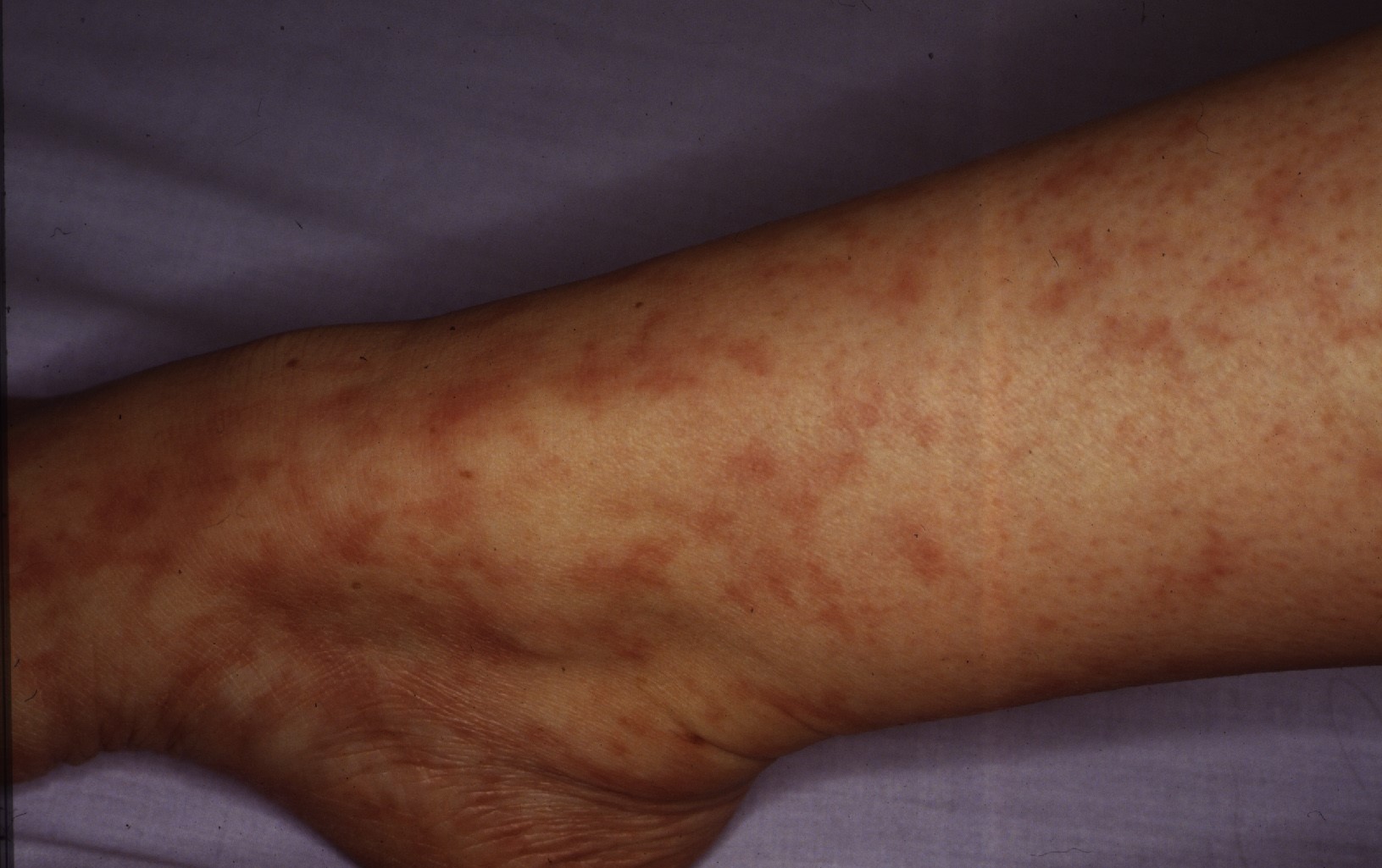
Image for DWII on lymphocytic thrombophilic arteritis
Image from DermNetNZ.org.

Five years later, Lee et al described 5 women (mean age, 25 years; age range, 20-34 years) with persistent, slowly progressive, patchy and reticular hyperpigmentation associated with livedo racemose, predominantly affecting the lower extremities. Most patients had only "subtle palpable subcutaneous indurations, with 1 patient having more prominent nodularity and papules." Biopsies revealed infiltration of the muscular vessel wall by inflammatory cells, affecting small arteries of the dermosubcutaneous junction or superficial subcutis. At least 90% of the infiltrate were lymphocytes with an admixture of histiocytes. Neutrophils and eosinophils were absent or scant, although one image clearly demonstrates intraluminal nuclear dust. A concentric fibrin ring involving the entire periphery of the lumina of affected vessels was present in all the patients, reminiscent of a thrombophilic state (which the authors thought could represent a localized thrombophilia triggered by lymphocytic endovasculitis). Laboratory studies demonstrated serum antiphospholipid antibodies in 4 patients, although none fulfilled criteria for having the antiphospholipid syndrome. One of these patients had a heterozygous mutation of the factor V Leiden gene. The authors believed that the vasculitic process in their patients was identical to those with MA as described by Fein et al. The authors concluded that the term "lymphocytic thrombophilic arteritis" best reflected the histologic features combining lymphocytic vascular inflammation with changes representing a thrombophilic endovasculitis. (3)

In an accompanying editorial, LeBoit opines: "Whether one calls this emerging condition macular arteritis (which invites confusion with a retinal condition) or lymphocytic thrombophilic arteritis, as Lee et al propose, it seems likely that there is now a condition in which lymphocytic vasculitis, in this case arteriolitis, is a characteristic finding." (4)

Macarenco et al reported 2 cases of biopsy-proven cutaneous lymphocytic arteritis. A 19-year-old white woman presented with hyperpigmented macules (and a few erythematous macules and papules) of her legs, that had been mildly painful a few months earlier. Other than an elevated ESR, her serologic evaluation was normal. The patient declined treatment and her lesions persisted over the next year. The second patient was a 26-year-old woman, with a prior history of deep vein thrombosis (with a heterozygous prothrombin mutation and wild type Factor V Leiden and plasminogen activator type-1 genes) presented with a 1-week history of leg pain and swelling, rash, fever, and pleuritic and epigastric pain. Serologic studies were negative, as were her Doppler and chest CT studies. A prompt remission was observed with a prednisone taper. Based on these cases and their literature review, the authors suggested that MA, LTA, and CPAN represent a spectrum of illness: "MA and LTA can be considered latent, non-nodule forming variants of CPAN [cutaneous polyarteritis nodosa], exhibiting less severe inflammatory destruction of the vessel wall and less pronounced neoangiogenesis (a histologic correlate with nodule formation). Increasing disease severity moving from MA to LTA to CPAN to systemic PAN is likely manifested by the accumulation of more numerous and varied types of vasculitic lesions (e.g., nodules, ulcers, foot drop) and acquisition of autoimmune serology (eg, ANA, RF, APA, and/or ANCA)." (5) In their systematic review of LTA, Vakili et al propose that MA and LTA are identical entities, and suggest that just the term LTA be utilized in instead of MA or MLA. (6)

Therapy for LTA has been mostly supportive, as no cases have been reported to progress to systemic vasculitis. Corticosteroids (topical or systemic), anticoagulants, antibiotics, and narrow band UVB have been utilized with little or no benefit. (7)

To date, there have now been approximately 50 reported cases of LTA and the controversy as to whether this entity is part of the CPAN spectrum remains unsettled. Buffiere-Morgado et al performed a retrospective study of 35 patients to assess the frequency of clinical and histologic features of MLA in patients with cPAN. All 35 patients had an infiltrated livedo, nodules, or both. Ulceration was rare. Erythematous or pigmented lesions were present in 54% of patients. Predominantly lymphocytic arteritis, a paucity of neutrophils, concentric fibrin ring, and absence of internal lamina elastic disruption were present in 60%, 20%, 18%, and 23% of patients, respectively. Median follow-up was 11 years. None of the patients had systemic involvement, and 57% had a complete remission. The incidence of complete remission was not different between patients having a predominant lymphocyte infiltrate or few neutrophils. The authors concluded that their data do not favor the classification of CPAN and MLA as distinct entities. (8) Other authors disagree. Kelly et al performed a cross-sectional study of 17 patients with LTA and 13 patients with CPAN. "Clinically, cases of LTA were distinguished by a more widespread pattern of livedo racemosa, which was non-infiltrated and asymptomatic. In contrast, cPAN was associated with localized starburst livedo, purpura, and episodic features including nodules, pain, and large inflammatory ulcers. When patients were separated according to the presence (>5%) or paucity (<5%) of neutrophils on blinded histology review, they had distinct clinical features and differences in disease course." The authors assert that their data supports the classification of LTA and CPAN as separate entities, underscoring the importance of clinical-pathologic correlation in distinguishing these disorders. (9)

In my estimation, the quandary of whether LTA falls within the spectrum of CPAN will not be answerable until we completely understand the pathogenesis of each disorder. Regardless, there is now ample literature to convince me that lymphocytic vasculitis is rare but real. Until such time when authorities definitively comprehend the etiology and nosology of LTA, the most prudent approach is to reassure the patient that this is likely a benign process, with an excellent prognosis, while following the patient carefully, being certain that there is no systemic vasculitis. 

Point to Remember: Lymphocytic thrombocytic vasculitis is a rare disorder with a good prognosis. Whether this falls under the spectrum of cutaneous polyarteritis nodosa, or is a disease sui generis, will be determined when the etiology of these diseases is ascertained. 

Our expert's viewpoint

Jason B. Lee, MD, FAAD
Clinical Vice Chair
Director, Jefferson Dermatopathology Center
Director, Residency and Dermatopathology Fellowship
Director, Jefferson Pigmented Lesion Clinic
Jefferson University Hospitals

Whether one believes macular arteritis and the more recent designation lymphocytic thrombophilic arteritis (LTA) is a distinct entity or it represents a spectrum of cutaneous polyarteritis nodosa (cPAN) will depend on which body of evidence one chooses to believe, as there is literature that supports either position. The rarity of both diseases makes it difficult to have a firm conviction about their nature. One must rely on limited or non-existent experience with the diseases and the scant body of evidence in the literature. 

Those who believe that LTA is a distinct entity focus on the clinical and histopathological differences when compared to cPAN, a view shared by LeBoit in his editorial where he affirms the existence of lymphocytic vasculitis pointing out that LTA is a prime example of one. (2-4, 9) Those who have a contrary view argue that the clinical and histopathological findings are not sufficiently distinct and that they can be encountered in the spectrum of clinical and histopathological presentation of cPAN, lumping the rarer LTA into a more established disease. According to these authors, the predominance of lymphocytes in cases reported as LTA represents late or reparative stage of cPAN. (5,8)

Neither point of view is supported by strong scientific body evidence, supported only by case reports and limited number of small studies. First is the issue of lymphocytic vasculitis, that is, whether lymphocytes may serve as the primary mediator of vasculitis. While neutrophilic vasculitis is well-established accounting for a majority of the vasculitis encountered in the skin, lymphocytic vasculitis is exceedingly rare. There is even debate whether this type of vasculitis even exists at all, some asserting that it merely represents an epiphenomenon or a secondary finding and not part of a primary pathologic process. I am part of the group who share this view for I have yet to encounter a convincing case of lymphocytic vasculitis as a dermatopathologist. In the few instances over the last 25 years where I have encountered the histopathologic changes described in LTA, I have signed them out as medium vessel vasculitis consistent with or favoring polyarteritis nodosa. Clinically, they were confirmed to be either a thrombotic vasculopathy of hypercoagulable state or cPAN. From my vantage, there is insufficient evidence to conclude that LTA is a clinically and histologically distinct entity. Late-stage or subacute forms of cPAN may account for the clinical and histopathological findings described in LTA. Until more convincing evidence emerges, I consider LTA as either a subset of cPAN or thrombotic vasculopathy rather than a disease sui generis.

  1. Watts RA, Robson J. Introduction, epidemiology, and classification of vasculitis. Best Pract Res Clin Rheumatol 2018; 32: 3-20.

  2. Fein H, Sheth AP, Mutasim DF. Cutaneous arteritis presenting with hyperpigmented macules: Macular arteritis. J Am Acad Dermatol 2003; 49: 519-522. 

  3. Lee JS, Kossard S, McGrath MA. Lymphocytic thrombophilic arteritis: A newly described medium-sized arteritis of the skin. Arch Dermatol 2008; 144: 1175-1182.

  4. LeBoit PE. The enigma of lymphocytic vasculitis. Arch Dermatol 2008; 144: 1215-1216.

  5. Macarenco RS, Galan A, Simoni PM, Macarenco AC, et al. Cutaneous lymphocytic thrombophilic (macular) arteritis: A distinct entity or an indolent (reparative) stage of cutaneous polyarteritis nodosa? A report of 2 cases of cutaneous arteritis and review of the literature. Am J Dermatopathol 2013; 35: 213-219. 

  6. Vakili S, Zampella JG, Kwatra SG, Blanck J, Loss M. Lymphocytic thrombophilic arteritis: A review. J Clin Rheumatol 2019; 25: 147-152. 

  7. Zampella JG, Vakili S, Doig S, Girardi N, et al. Macular lymphocytic arteritis: Clinical-pathologic correlation of a rare vasculitis. JAAD Case Rep 2017; 3: 116-120.

  8. Buffiere-Morgado A, Battistella M, Vignon-Pennamen MD, de Masson A, et al. Relationship between cutaneous polyarteritis nodosa (cPAN) and macular lymphocytic arteritis (MLA): Blinded histologic assessment of 35 cPAN cases. J Am Acad Dermatol 2015; 73: 1013-1020. 

  9. Kelly RI, Wee E, Balta S, Williams RA. Lymphocytic arteritis and cutaneous polyarteritis nodosa: Clinicopathologic comparison with blinded histologic assessment. J Am Acad Dermatol 2020 Feb 7 [Online ahead of print].


All content found on Dermatology World Insights and Inquiries, including: text, images, video, audio, or other formats, were created for informational purposes only. The content represents the opinions of the authors and should not be interpreted as the official AAD position on any topic addressed. It is not intended to be a substitute for professional medical advice, diagnosis, or treatment.

DW Insights and Inquiries archive

Explore hundreds of Dermatology World Insights and Inquiriesarticles by clinical area, specific condition, or medical journal source.


Sent from my iPhone

Benjamin Hidalgo-Matlock
Skin Care Physicians of Costa Rica

Clinica Victoria en San Pedro: 4000-1054
Momentum Escazu: 2101-9574

Please excuse the shortness of this message, as it has been sent from
a mobile device.

Monday, May 09, 2022

Paninflammation Value in HS

The pan-immune-inflammation value (PIV) is a recently proposed scoring system that includes all immune-inflammatory cells in the peripheral blood count (neutrophil count × platelet count × monocyte count)/lymphocyte count).

Blood Collection-Based Systemic Inflammation Biomarkers for Patients With Hidradenitis Suppurativa

Journal of the European Academy of Dermatology and Venereology: JEADV


TAKE-HOME MESSAGE


Abstract 

BACKGROUND

Hidradenitis suppurativa (HS) is a relatively common chronic inflammatory condition of intertriginous skin. In recent years, the neutrophil/lymphocyte ratio (NLR), platelet/lymphocyte ratio (PLR), monocyte/lymphocyte ratio (MLR), and platelet/neutrophil ratio (PLR) have been shown to be indicators of systemic inflammation correlating with severity of inflammatory conditions.

OBJECTIVES

We aimed to analyse for the first time systemic inflammation biomarkers also including the pan-immune-inflammation value (PIV) and the systemic immune-inflammation index (SII) in HS patients and controls.

METHODS

This study retrospectively investigated clinical and laboratory data of 142 patients with HS. Moreover, a sex-age-matched healthy control group was included. The severity of HS was routinely assessed by the Hurley staging, the mHSS, and the SAHS score. All inflammation-based biomarkers were calculated from absolute values of complete blood counts. Receiver operating characteristics analyses, including Youden index, were performed in order to determine optimal cut-off values and test performance.

RESULTS

Whereas PIV and SII were significantly higher in HS patients, PLR, MLR, and PNR were significantly lower in HS patients when compared to controls. Almost all inflammation-based biomarkers significantly correlated with disease severity. However, PIV was the only test that was significantly associated with HS severity as indicated by a Youden index of 0.56 (associated criterion: 756.4; AUC: 0.79, P < 0.0001).

CONCLUSIONS

Although all systemic inflammation-based biomarkers investigated are more or less associated with HS severity, the PIV appears to have the best performance in this regard. It may be employed in adjunction with the clinical scores for treatment decision making or clinical trial assessments.


Journal of the European Academy of Dermatology and Venereology: JEADV
Complete blood collection-based systemic inflammation biomarkers for patients with hidradenitis suppurativa
J Eur Acad Dermatol Venereol 2022 Apr 24;[EPub Ahead of Print], T Gambichler, S Hessam, P Cramer, N Abu Rached, G Bechara 

Sent from my iPhone

Benjamin Hidalgo-Matlock
Skin Care Physicians of Costa Rica

Clinica Victoria en San Pedro: 4000-1054
Momentum Escazu: 2101-9574

Please excuse the shortness of this message, as it has been sent from
a mobile device.

Sunday, May 08, 2022

Topical treatment pyogenic granulomas!

Topical treatment of pyogenic granulomas in a pediatric population: A single-institution retrospective review

Key words

To the Editor: Pyogenic granulomas (PG) are common benign vascular proliferations of the skin and mucous membranes. Because of their propensity to grow and bleed, surgical management is favored, but it can be costly, traumatic, result in scarring, or require an in-office or operative suite setting. Noninvasive treatment modalities include topical beta-blockers, imiquimod, and corticosteroids. By promoting vasoconstriction and downregulating proangiogenic factors such as vascular endothelial growth factor A, matrix metalloproteinase 1, and interleukin 6, topical corticosteroids potentially target several key pathways implicated in the PG pathogenesis.,Here, we expand the limited data supporting the use and efficacy of high potency topical corticosteroids for the treatment of PG and analyze the use of topical and procedural interventions.

Following IRB approval, data were collected retrospectively using the International Statistical Classification of Diseases, Tenth Revision code L98.0 for patients diagnosed with PG at the Boston Children's Hospital from January 1, 2019, to September 13, 2021. Cases with incongruent biopsy results or without follow-up were excluded. Data including demographics, treatment, complications, clinical characteristics, and outcomes were recorded. Statistical analyses were completed using STATA. Statistical significance was set to P < .05.

Ninety-eight patients were included (mean age, 9.8 years; range, 0.5-22 years) (Table I). Sixty-three patients were managed surgically; 50 with in-office shave removal plus electrocautery and 13 with excision. Regrowth occurred after shave removal in 4 patients and 1 patient on filgrastim (with >50 eruptive PG) after excision (Table II). Surgical site infection occurred in 1 patient; 4 patients followed up with concerns about their scar after the procedure.

Table IPatient demographics and pyogenic granuloma characteristics

PG, Pyogenic granuloma.

∗ Two-sided t test was performed for continuous variables and χ2 test for categorical variables. Significance was set at P < .05.

† Intrinsic or secondary to systemic medical therapy.

‡ N = 79.

§ For patients with >1 lesion, each area of involvement was counted individually.

Table IIPatient demographics, pyogenic granuloma characteristics, and clinical outcomes by treatment modality

PG, Pyogenic granuloma.

∗ Treated with both modalities either simultaneously or sequentially.

Thirty-five patients were treated noninvasively; 31 on initial presentation and 4 after initial procedural intervention. Patients who were managed topically were younger (P = .013), and their PG size was smaller (P = .006) (Table II). Fourteen patients received a class I topical corticosteroid (clobetasol [86%], halobetasol [7%], and betamethasone [7%]) alone, applied twice daily under occlusion for a median of 5 weeks (range, 5 days-2 years). Sixteen patients received topical timolol (0.5% ophthalmic gel-forming solution) alone for a median of 6 weeks (range, 8 days-1.6 years). Partial or complete resolution with topical timolol or topical corticosteroid was 50%. Patients managed with topical corticosteroid versus topical timolol underwent subsequent excision in 43% and 56% of cases, respectively (P = .464). Five patients were treated with both topical modalities (Table II). Lesions ≤4 mm responded favorably to the topical therapy. No adverse effects of topical therapy were reported.

There are no standard guidelines for the management of PG. However, both topical corticosteroids and timolol have excellent safety profiles and are widely used for numerous pediatric skin conditions. Noninvasive management may be favored for younger patients, poor surgical candidates, those with eruptive lesions, or those with small lesions in cosmetically sensitive areas. The COVID-19 pandemic exacerbated barriers in access to care and minimized in-person appointments for many specialties; thus, topical treatment may benefit patients with limited access to live care or those awaiting procedural intervention. Additionally, topical treatment is less suitable for actively bleeding PG, lesions with unclear diagnosis, or patients seeking a rapid cure. Patients and parents should be counseled that topical therapy may not work, and that they may elect to pursue excision later.

Topical treatment may be continued as long as improvement is noted; if PG fails to improve, or if adverse effects are noted, an alternative topical agent or procedure may be pursued. Lesions that exhibit concerning features should be evaluated and may require a biopsy as infectious, malignant, or other vasoproliferative lesions remain on the differential. In this study, patients were not randomized to treatment modalities; thus, the success of the topical treatment is impacted by selection bias whereby experienced clinicians selected patients most amenable to the topical treatment. Although limited by its retrospective design, small sample size, and the use of a single center, our study supports a potential role for the topical management of PG.


Sent from my iPhone

Benjamin Hidalgo-Matlock
Skin Care Physicians of Costa Rica

Clinica Victoria en San Pedro: 4000-1054
Momentum Escazu: 2101-9574

Please excuse the shortness of this message, as it has been sent from
a mobile device.

Tuesday, May 03, 2022

Bleach baths for atopic dermatitis

Bleach baths for atopic dermatitis

A systematic review and meta-analysis including unpublished data, Bayesian interpretation, and GRADE
Published:March 29, 2022DOI:https://doi.org/10.1016/j.anai.2022.03.024

Abstract

Background

Bleach bathing is frequently recommended to treat atopic dermatitis (AD), but its efficacy and safety are uncertain.

Objective

To systematically synthesize randomized controlled trials (RCTs) addressing bleach baths for AD.

Methods

We searched MEDLINE, EMBASE, CENTRAL, and GREAT from inception to December 29, 2021, for RCTs assigning patients with AD to bleach vs no bleach baths. Paired reviewers independently and in duplicate screened records, extracted data, and assessed risk of bias (Cochrane version 2) and GRADE quality of evidence. We obtained unpublished data, harmonized individual patient data and did Frequentist and Bayesian random-effects meta-analyses.

Results

There were 10 RCTs that enrolled 307 participants (median of mean age 7.2 years, Eczema Area Severity Index baseline mean of means 27.57 [median SD, 10.74]) for a median of 6 weeks (range, 4-10). We confirmed that other trials registered globally were terminated. Bleach baths probably improve AD severity (22% vs 32% improved Eczema Area Severity Index by 50% [ratio of means 0.78, 95% credible interval 0.59-0.99]; moderate certainty) and may slightly reduce skin Staphylococcal aureus colonization (risk ratio, 0.89 [95% confidence interval, 0.73-1.09]; low certainty). Adverse events, mostly dry skin and irritation, along with itch, patient-reported disease severity, sleep quality, quality of life, and risk of AD flares were not clearly different between groups and of low to very low certainty.

Conclusion

In patients with moderate-to-severe AD, bleach baths probably improve clinician-reported severity by a relative 22%. One in 10 will likely improve severity by 50%. Changes in other patient-important outcomes are uncertain. These findings support optimal eczema care and the need for additional large clinical trials.

Trial Registration

PROSPERO Identifier: CRD42021238486.

Sent from my iPhone

Benjamin Hidalgo-Matlock
Skin Care Physicians of Costa Rica

Clinica Victoria en San Pedro: 4000-1054
Momentum Escazu: 2101-9574

Please excuse the shortness of this message, as it has been sent from
a mobile device.

Sunday, May 01, 2022

Comparing the frequency of isotretinoin-induced hair loss at <0.5-mg/kg/d versus ≥0.5-mg/kg/d dosing in acne patients: A systematic review

Systematic reviews/meta-analyses| Volume 6, P125-142, March 01, 2022

Comparing the frequency of isotretinoin-induced hair loss at <0.5-mg/kg/d versus ≥0.5-mg/kg/d dosing in acne patients: A systematic review

Open AccessPublished:February 10, 2022DOI:https://doi.org/10.1016/j.jdin.2022.01.002


Over 1 million isotretinoin prescriptions are authorized in the United States per year. An insight into the frequency, dose dependency, timing, and reversibility of hair loss associated with isotretinoin treatment for acne vulgaris could help guide dosing regimens and patient counseling. The objective of this systematic review was to assess the frequency of hair loss in patients with acne vulgaris on <0.5 mg/kg/d daily doses of isotretinoin versus the frequency of hair loss in patients with acne vulgaris on ≥0.5 mg/kg/d daily doses of isotretinoin.
An Embase and MEDLINE search was conducted on July 15, 2020, in accordance with the Preferred Reporting Items for Systematic Reviews and Meta-Analyses guidelines. The review focused on acne vulgaris patients. The treatment of acne vulgaris is the most common use of isotretinoin, and the population is typically younger and with fewer comorbidities.
Twenty-two studies reported hair loss with oral isotretinoin treatment. A frequency analysis suggested that patients with acne vulgaris on <0.5 mg/kg/d of isotretinoin experienced hair loss at a frequency of 3.2% (n = 18/565) compared with those on ≥0.5 mg/kg/d, who experienced hair loss at a frequency of 5.7% (n = 192/3375). Inferential statistics were not possible.
Physicians should consider counseling patients about the risk of telogen effluvium prior to drug initiation, as is commonly done for other side effects. The potential trend of increased hair loss frequency at a higher daily dosing warrants further investigation using higher-quality research.

The Science of Clean Skin Care and the Clean Beauty Movement

Cosmeceutical Critique

The Science of Clean Skin Care and the Clean Beauty Movement

Leslie S. Baumann, MD

April 29, 2022

As the clean beauty movement is gaining momentum, it has become challenging to differentiate between science and marketing hype. I see numerous social media posts, blogs, and magazine articles about toxic skin care ingredients, while more patients are asking their dermatologists about clean beauty products. So, I decided it was time to dissect the issues and figure out what "clean" really means to me.

Dr Leslie Baumann

The problem is that no one agrees on a clean ingredient standard for beauty products. Many companies, like Target, Walgreens/Boots, Sephora, Neiman Marcus, Whole Foods, and Ulta, have their own varying clean standards. Even Allure magazine has a "Clean Best of Beauty" seal. California has Proposition 65, otherwise known as the Safe Drinking Water and Toxic Enforcement Act of 1986, which contains a list of banned chemicals "known to the state to cause cancer or reproductive toxicity." In January 2021, Hawai'i law prohibited the sale of oxybenzone and octinoxate in sunscreens in response to scientific studies showing that these ingredients "are toxic to corals and other marine life." The Environmental Working Group (EWG) rates the safety of ingredients based on carcinogenicity, developmental and reproductive toxicity, allergenicity, and immunotoxicity. The Cosmetic Ingredient Review (CIR), funded by the Personal Care Products Council, consists of a seven-member steering committee that has at least one dermatologist representing the American Academy of Dermatology and a toxicologist representing the Society of Toxicology. The CIR publishes detailed reviews of ingredients that can be easily found on PubMed and Google Scholar and closely reviews animal and human data and reports on safety and contact dermatitis risk.

Which Clean Beauty Standard Is Best?

I reviewed most of the various standards, clean seals, laws, and safety reports and found significant discrepancies resulting from misunderstandings of the science, lack of depth in the scientific evaluations, lumping of ingredients into a larger category, or lack of data. The most salient cause of misinformation and confusion seems to be hyperbolic claims by the media and clean beauty advocates who do not understand the basic science.

When I conducted a survey of cosmetic chemists on my LinkedIn account, most of the chemists stated that " 'Clean Beauty' is a marketing term, more than a scientific term." None of the chemists could give an exact definition of clean beauty. However, I thought I needed a good answer for my patients and for doctors who want to use and recommend "clean skin care brands."

A Dermatologist's Approach to Develop a Clean Beauty Standard

Many of the standards combine all of the following into the "clean" designation: nontoxic to the environment (both the production process and the resulting ingredient), nontoxic to marine life and coral, cruelty-free (not tested on animals), hypoallergenic, lacking in known health risks (carcinogenicity, reproductive toxicity), vegan, and gluten free. As a dermatologist, I am a splitter more than a lumper, so I prefer that "clean" be split into categories to make it easier to understand. With that in mind, I will focus on clean beauty ingredients only as they pertain to health: carcinogenicity, endocrine effects, nephrotoxicity, hepatotoxicity, immunotoxicity, etc. This discussion will not consider environmental effects, reproductive toxicity (some ingredients may decrease fertility, which is beyond the scope of this article), ingredient sources, and sustainability, animal testing, or human rights violations during production. Those issues are important, of course, but for clarity and simplicity, we will focus on the health risks of skin care ingredients.

In this month's column, I will focus on a few ingredients and will continue the discussion in subsequent columns. Please note that commercial standards such as Target standards are based on the product type (e.g., cleansers, sunscreens, or moisturizers). So, when I mention an ingredient not allowed by certain company standards, note that it can vary by product type. My comments pertain mostly to facial moisturizers and facial serums to try and simplify the information. The Good Face Project has a complete list of standards by product type, which I recommend as a resource if you want more detailed information.

Are Ethanolamines Safe or Toxic in Cosmetics?

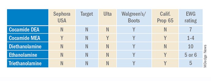
Ethanolamines are common ingredients in surfactants, fragrances, and emulsifying agents and include cocamide diethanolamine (DEA), cocamide monoethanolamine (MEA), and triethanolamine (TEA). Cocamide DEA, lauramide DEA, linoleamide DEA, and oleamide DEA are fatty acid diethanolamides that may contain 4% to 33% diethanolamine.1 A Google search of toxic ingredients in beauty products consistently identifies ethanolamines among such offending product constituents. Table 1 reveals that ethanolamines are excluded from some standards and included in others (N = not allowed or restricted by amount used and Y = allowed with no restrictions). As you can see, the standards don't correspond to the EWG rating of the ingredients, which ranges from 1 (low hazard) to 10 (high hazard).


Why Are Ethanolamines Sometimes Considered Safe and Sometimes Not?

Ethanolamines are reputed to be allergenic, but as we know as dermatologists, that does not mean that everyone will react to them. (In my opinion, allergenicity is a separate issue than the clean issue.) One study showed that TEA in 2.5% petrolatum had a 0.4% positive patch test rate in humans, which was thought to be related more to irritation than allergenicity.2 Cocamide DEA allergy is seen in those with hand dermatitis resulting from hand cleansers but is more commonly seen in metal workers.3 For this reason, these ethanolamines are usually found in rinse-off products to decrease exposure time. But there are many irritating ingredients not banned by Target, Sephora, and Ulta, so why does ethanolamine end up on toxic ingredient lists?

First, there is the issue of oral studies in animals. Oral forms of some ethanolamines have shown mild toxicity in rats, but topical forms have not been demonstrated to cause mutagenicity.1

For this reason, ethanolamines in their native form are considered safe.

The main issue with ethanolamines is that, when they are formulated with ingredients that break down into nitrogen, such as certain preservatives, the combination forms nitrosamines, such as N-nitrosodiethylamine (NDEA), which are carcinogenic.4 The European Commission prohibits DEA in cosmetics based on concerns about formation of these carcinogenic nitrosamines. Some standards limit ethanolamines to rinse-off products.5 The CIR panel concluded that diethanolamine and its 16 salts are safe if they are not used in cosmetic products in which N-nitroso compounds can be formed and that TEA and TEA-related compounds are safe if they are not used in cosmetic products in which N-nitroso compounds can be formed.6,7 The FDA states that there is no reason for consumers to be alarmed based on the use of DEA in cosmetics.

The safety issues surrounding the use of ethanolamines in a skin care routine illustrate an important point: Every single product in the skin care routine should be compatible with the other products in the regimen. Using ethanolamines in a rinse-off product is one solution, as is ensuring that no other products in the skin care routine contain N-nitroso compounds that can combine with ethanolamines to form nitrosamines.

Are Natural Products Safer?

Natural products are not necessarily any safer than synthetic products. Considering ethanolamines as the example here, note that cocamide DEA is an ethanolamine derived from coconut. It is often found in "green" or "natural" skin care products.9 It can still combine with N-nitroso compounds to form carcinogenic nitrosamines.

What Is the Bottom Line? Are Ethanolamines Safe in Cosmetics?

For now, if a patient asks if ethanolamine is safe in skin care, my answer would be yes, so long as the following is true:

  • It is in a rinse-off product.

  • The patient is not allergic to it.

  • They do not have hand dermatitis.

  • Their skin care routine does not include nitrogen-containing compounds like N-nitrosodiethanolamine (NDELA) or NDEA.

Conclusion

This column uses ethanolamines as an example to show the disparity in clean standards in the cosmetic industry. As you can see, there are multiple factors to consider. I will begin including clean information in my cosmeceutical critique columns to address some of these issues.

Baumann is a private practice dermatologist, researcher, author, and entrepreneur who practices in Miami. She founded the Cosmetic Dermatology Center at the University of Miami in 1997. Baumann has written two textbooks and a New York Times Best Sellers book for consumers. Baumann has received funding for advisory boards and/or clinical research trials from Allergan, Galderma, Revance, Evolus, and Burt's Bees. She is the CEO of Skin Type Solutions Inc., a company that independently tests skin care products and makes recommendations to physicians on which skin care technologies are best. Write to her at dermnews@mdedge.com.

References

1. Cocamide DE. J Am Coll Toxicol. 1986;5(5).

2. Lessmann H et al. Contact Dermatitis. 2009 May;60(5):243-55.

3. Aalto-Korte K et al. 2014 Mar;70(3):169-74.

4. Kraeling ME et al. Food Chem Toxicol. 2004 Oct;42(10):1553-61.

5. Fiume MM et al. Int J Toxicol. 2015 Sep;34(2 Suppl):84S-98S.

6. Fiume MM.. Int J Toxicol. 2017 Sep/Oct;36(5_suppl2):89S-110S.

7. Fiume MM et al. Int J Toxicol. 2013 May-Jun;32(3 Suppl):59S-83S.

8. U.S. Food & Drug Administration. Diethanolamine. https://www.fda.gov/cosmetics/cosmetic-ingredients/diethanolamine. Accessed Feb. 12, 2022.

9. Aryanti N et al. IOP Conference Series: Materials Science and Engineering 2021 Feb 1 (Vol. 1053, No. 1, p. 012066). IOP Publishing.

This article originally appeared on MDedge.com, part of the Medscape Professional Network.