Dermatología en Costa Rica

Thursday, July 30, 2020

BP topical treatment

Superpotent Topical Corticosteroid Therapy in Bullous Pemphigoid

The British Journal of Dermatology


TAKE-HOME MESSAGE


Abstract

Superpotent topical corticosteroids (TCS) have been demonstrated to be more effective and safer than high doses of oral corticosteroids to treat extensive bullous pemphigoid (BP).1 They are proposed as first‐line treatment for BP in the Cochrane review2 and in European guidelines.3 In France, TCS are the recommended first‐line treatment for BP, regardless of disease severity.4 The objectives of this study were to determine whether TCS alone were actually used as first‐line therapy and could be successfully continued during the first year of treatment in routine practice, and to estimate the related healthcare costs.

The British Journal of Dermatology
Feasibility and Healthcare Costs of Superpotent Topical Corticosteroid Therapy in Bullous Pemphigoid: A Prospective, Observational Study in an Academic Centre in France
Br J Dermatol 2020 Apr 22;[EPub Ahead of Print], A Clapé, C Muller, J Plée, M Viguier, C Vanhaecke, P Bernard 


Sent from my iPhone

Benjamin Hidalgo-Matlock
Skin Care Physicians of Costa Rica

Clinica Victoria en San Pedro: 4000-1054
Momentum Escazu: 2101-9574

Please excuse the shortness of this message, as it has been sent from
a mobile device.

Wednesday, July 29, 2020

Síndrome oreja roja...

WHY YOU SHOULD HEAR ABOUT THE RED EAR SYNDROME


DII small banner

By Warren R. Heymann, MD
July 22, 2020
Vol. 2, No. 29

Is it my imagination that the "by the way" comments at the presumed end of a patient encounter cause the most angst? Usually, it's something like "Oh, I forgot to mention that I'm losing my hair!" Recently, at the conclusion of an extended visit, a middle-aged woman stated that periodically, her left ear would become fiery red for several hours, accompanied by a burning sensation. There was no evidence of disease that day — my quick differential of a contact dermatitis, Pseudomonas cellulitis, relapsing polychondritis, or chilblains, were not pertinent. All I could do was punt — "Why don't you see me when you get one of these episodes? I'll take a look, and assess it at that time" (while thinking that I have no idea what she has)?

Perhaps my patient is experiencing the red ear syndrome (RES); I was not familiar with this entity until I read the article by Cinats and Haber. (1) Wollina contends that RES is one of a triad of orphan disorders that dermatologists should recognize, along with the red scrotum and red scalp syndromes. (2)

RES was first described by Lance in 1996, detailing the features of a dozen case reports. The following is the first paragraph of his discussion: "What are the characteristics of the red ear syndrome? It is commonly unilateral and implies discomfort or burning pain as well as a change in color. The color change may involve the adjacent part of the cheek. The pain may radiate to the forehead (V-1), occiput (C-2), or a strip behind or below the mandible (C-3). It is often brought on by exercise, exposure to heat, touch, chewing, or neck movement." Prior to publication of this article, Dr. Lance stated that he presented three of these patients to the Australian Association of Neurologists a paper entitled "The Mystery of One Red Ear." (3)

Illustration for DWII on RES
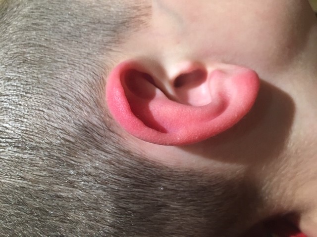
Photo courtesy of Richard Haber, MD

Nearly a quarter century later, RES remains mysterious. Most of the information about RES has been in the neurology and otolaryngology literature, so it is not surprising that dermatologists may not be familiar with the entity. There is considerable debate about the classification, pathophysiology, and treatment of this (possibly not so) rare disorder.

According to Lambru et al, RES episodes are characterized by mostly unilateral, or occasionally bilateral, attacks of paroxysmal burning sensations and reddening of the external ear. This includes the earlobe [distinguishing RES from relapsing polychondritis]. The duration of these episodes ranges from a few seconds to several hours. Attacks occur with a frequency ranging from several a day to a few per year; they may occur spontaneously or be triggered by rubbing or touching the ear, heat or cold, chewing, brushing of the hair, neck movements, or exertion. Early-onset idiopathic (primary) RES seems to be associated with migraine, whereas late-onset secondary forms of RES occur with trigeminal autonomic cephalalgias (TACs), upper cervical spine disorders, or temporo-mandibular joint dysfunction. (Please see below for their proposed diagnostic criteria primary RES.) RES is mostly refractory to medical treatments, although some migraine preventative treatments have shown moderate benefit mainly in patients with migraine-related attacks. (4) Some recent therapeutic suggestions include use of botulinum toxin (5), indomethacin (6), or propranolol. (7) A partial list of other treatments includes topical anesthetics, oral tricyclic antidepressants, and gabapentin. (1) Physical cooling of the skin may be worthwhile. (2)

The pathophysiology of RES remains to be determined. Chan et al state: "The vasomotor control of the skin of the ear is dependent on sympathetic vasoconstrictor tone as opposed to the forehead and cheek skin, which is predominantly influenced by the facial parasympathetic system as seen in trigeminal autonomic cephalalgias (TACs). It seems unlikely, therefore, that the trigemino‐parasympathetic reflex accounts for the erythema of the ear, although it is possible that an imbalance between the parasympathetic and sympathetic system may in turn result in inhibition of sympathetic tone in the ear, giving rise to vasodilation and the red ear phenomenon." It is also conceivable that irritation of cervical nerve roots or the mandibular trigeminal nerve could activate these autonomic circuits. (6) Cinats and Haber presented the case of an 11-year-old boy with daily episodes of RES accompanied by erythema and swelling of his hands and feet. Based on these findings, they suggest that RES may represent auricular erythromelalgia. The authors propose a new classification of RES including primary forms (divided into headache or erythromelalgia types) and secondary RES.

One of the joys of reading widely is experiencing an "aha" moment of recognizing the relevancy of an unknown disorder (to me) to an existing patient's problem. When my patient returns, I will strongly consider the diagnosis of the red ear syndrome (RES), thanks to the publication by Cinats and Haber.

Point to Remember: The red ear syndrome may be a primary or secondary disorder that displays symptoms similar to erythromelalgia. After ruling out other conditions that may cause auricular erythema, the diagnosis of RES should be considered — the best therapeutic approach may be multidisciplinary with assistance from neurologists and otolaryngologists.

Our expert's viewpoint

Richard M Haber MD, FRCPC
Professor of Medicine
University of Calgary

The main purpose of my article on pediatric red ear syndrome (RES) was to make this diagnosis better known to dermatologists, as most case reports of RES have appeared in the neurologic literature. I believe that some primary types of RES (such as my case in a pediatric patient) likely represent an erythromelalgia-type of RES (RES-ET). Cases such as my young boy with refractory painful erythema of his ears which could be associated with concurrent erythema of his hands and feet, strengthens the classification proposed in my article which includes RES-ET as a primary type of RES.

There is clearly a headache-associated primary type of RES (RES-HT) in which the episodes of auricular erythema affecting the whole ear including the lobule occur temporally. Interestingly, the headache and the red ear(s) may respond to treatment of the headache. The subset that is most confusing is patients with RES but with a history of headaches that are not concurrent with the auricular erythema. Whether this is a true subset of RES remains to be determined.

Although RES appears to be a rare diagnosis presenting to dermatologists, it is likely that more cases will present as a primary dermatologic complaint. When a Google search of "red ears" and "pain" is performed, the first diagnosis suggested is RES followed by relapsing polychondritis. It is therefore very likely that patients will present to a dermatologist asking about RES.

Treatment of RES, especially the RES-ET can be extremely challenging and I agree with Dr. Heymann's suggestion that difficult cases may best be managed with a multidisciplinary approach with assistance from neurologists and otolaryngologists but could also require a referral for assessment in specialized complex pain clinics similar to refractory cases of hand and foot erythromelalgia.

When it comes to ear syndromes, I suggest that dermatologists be well "red" and consider the diagnosis of RES.

Proposed diagnostic criteria for primary red ear syndrome (reference 4)

A. At least 20 attacks fulfilling criteria B-E
B. Episodes of external ear pain lasting up to 4 hours.
C. The ear pain has at least two of the following characteristics:
  -Burning quality
  -Unilateral location
  -Mild to moderate severity
  -Triggered by cutaneous or thermal stimulation of the ear.
D. The ear pain is accompanied by ipsilateral redness of the external ear.
E. Attacks occur with a frequency of ≥1 per day, although cases with lower frequency may occur.
F. Not attributed to another disorder.

  1. Cinats AK, Haber RM. Pediatric red ear syndrome: A case report of an erythromelalgia type and review of the literature. Pediatr Dermatol 36: 686-689.

  2. Wollina U. Three orphans one should know: Red scalp, red ear, and red scrotum syndrome. J Eur Acad Dermatol Venereol 2016; 30: e169-170.

  3. Lance JW. The red ear syndrome. Neurology 1996; 47: 617-620.

  4. Lambru G, Miller S, Matharu MS. The red ear syndrome. J Headache Pain 2013; 14: 83.

  5. Castellanos-Gonzalez M, Manueles F. Martos MG, Rodriguez MA. Successful treatment of red ear syndrome with botulinum toxin type A. Indian J Dermatol Venereol Leprol 2019; 85: 107-108.

  6. Chan TLH, Becker WJ, Jog M. Indomethacin-responsive idiopathic red ear syndrome: Case report and pathophysiology. Headache 2018; 58: 306-308.

  7. Hernández CR, Loza SM, López RL, del Mar García Romero. Red ear syndrome in a pediatric patient. Pediatr Emerg Care 2018; 34: e199-e200.


All content found on Dermatology World Insights and Inquiries, including: text, images, video, audio, or other formats, were created for informational purposes only. The content represents the opinions of the authors and should not be interpreted as the official AAD position on any topic addressed. It is not intended to be a substitute for professional medical advice, diagnosis, or treatment.

Monday, July 27, 2020

Obesidad y Covid

Journal Scan / Research · July 14, 2020

Association Between BMI and COVID-19 Hospitalization by Age

Obesity 


TAKE-HOME MESSAGE

Abstract 

OBJECTIVE

Obesity has been found to be a risk factor for hospitalization with COVID-19. We were interested in understanding whether patients hospitalized with COVID-19 differed in BMI at older versus younger ages, and if trends were independent of diabetes and hypertension.

METHODS

We performed a cross-sectional analysis of patients hospitalized with moderate to severe COVID-19 at Northwestern Memorial Hospital from March 19th , 2020 until April 4th , 2020. We compared patients hospitalized with COVID-19 above and below the age of 50, and to those hospitalized without COVID-19.

RESULTS

We found patients younger than 50 years of age hospitalized with COVID-19 without diabetes or hypertension had mean BMI greater than those older than 50 years of age, with BMI 43.1 (95%CI 34.5 - 51.7) kg/m2 vs 30.1 (95%CI 27.7 - 32.5) kg/m2 (p=0.02). Furthermore, BMI appears to inversely correlate with increasing age amongst patients hospitalized with COVID-19. We did not detect the same difference or trend for patients hospitalized without COVID-19.

CONCLUSION

We found younger patients (age <50 years) with COVID-19 had higher mean BMI than older patients with COVID-19, with and without diabetes and hypertension. This trend did not exist in patients without COVID-19 hospitalized during the same time-period.


Obesity 
Is BMI Higher in Younger Patients With COVID-19? Association Between BMI and COVID-19 Hospitalization by Age
Obesity 2020 Jul 01;[EPub Ahead of Print], A Bhasin, H Nam, C Yeh, J Lee, D Liebovitz, C Achenbach 

Sent from my iPhone

Benjamin Hidalgo-Matlock
Skin Care Physicians of Costa Rica

Clinica Victoria en San Pedro: 4000-1054
Momentum Escazu: 2101-9574

Please excuse the shortness of this message, as it has been sent from
a mobile device.

Rituximab y penfigo

Journal Scan / Research · July 22, 2020

Long-Term Outcomes of Rituximab Therapy in Pemphigus

Journal of the European Academy of Dermatology and Venereology: JEADV

TAKE-HOME MESSAGE


Abstract


BACKGROUND

Rituximab induces a rapid remission in most patients with pemphigus.

OBJECTIVE

Our aim was to assess the long-term efficacy of rituximab in this disease.

METHOD

We conducted a retrospective study of 59 patients with pemphigus treated with rituximab and observed over a median period of 104 months.

RESULTS

The rate of complete remission off therapy (CRoff) after the first rituximab cycle was 39%, increasing to 61% with additional rituximab courses. Long-term CRoff was achieved in 27% of patients. The recurrence rate after the first rituximab cycle was 63%, decreasing to approximately 40% with subsequent rituximab cycles. Median time to relapse after the first and subsequent rituximab cycles was 25 months. Renewed rituximab therapy reinduced complete remission in 94% of cases. Baseline anti-desmoglein antibody levels of ≤250 U/mL were significantly associated with the outcome of CRoff. In paired serum samples obtained before the first and six months after the last rituximab therapy, significant reductions of desmoglein-specific autoantibodies were observed. Patients relapsing after a complete remission induced by the first rituximab cycle were more likely to achieve CRoff than patients relapsing after a less favourable outcome and non-responders. There was no significant difference in age, sex, pemphigus subtype, rituximab dosing and disease duration between patients achieving CRoff and those not meeting this end point.

CONCLUSIONS

Lower desmoglein-specific antibody levels at baseline were predictive of CRoff. In patients receiving multiple rituximab cycles, complete remission after the first cycle was associated with a favourable long-term outcome. Repeated rituximab courses were highly effective for relapsed disease and improved the overall outcome.


Journal of the European Academy of Dermatology and Venereology: JEADV
Long-Term Outcomes of Rituximab Therapy in Pemphigus
J Eur Acad Dermatol Venereol 2020 May 04;[EPub Ahead of Print], I Shimanovich, T Baumann, E Schmidt, D Zillikens, CM Hammers 

Sent from my iPhone

Benjamin Hidalgo-Matlock
Skin Care Physicians of Costa Rica

Clinica Victoria en San Pedro: 4000-1054
Momentum Escazu: 2101-9574

Please excuse the shortness of this message, as it has been sent from
a mobile device.

Capecitabina oral

Journal Scan / Review · July 24, 2020

Capecitabine for the Treatment and Prevention of Actinic Keratoses, Squamous Cell Carcinoma, and Basal Cell Carcinoma

JAMA Dermatology

TAKE-HOME MESSAGE

Abstract


IMPORTANCE

Certain patient groups, such as solid organ transplant recipients (SOTRs), have a significantly increased risk of developing skin cancers. The chemotherapeutic drug capecitabine has been used off label as a chemopreventive modality to suppress the development of precancerous skin lesions and squamous cell carcinomas (SCCs).

OBJECTIVE

To systematically review published studies on the use of capecitabine for the treatment and prevention of precancerous and cancerous skin lesions, with a focus on cutaneous SCC.

EVIDENCE REVIEW

For this systematic review, a literature search was performed using the PubMed and Embase databases in December 2019 for all articles published between January 1, 1998, and December 31, 2019, using the search term capecitabine paired with each of the following terms: actinic keratosis, actinic keratoses, squamous cell carcinoma, and basal cell carcinoma. Articles on the use of capecitabine for the treatment and prevention of actinic keratoses (AKs), SCCs, and basal cell carcinomas (BCCs) were selected for inclusion.

FINDINGS

Sixteen publications met the criteria for inclusion, with 8 case reports describing the inflammation of AKs in patients with solid organ cancer treated with capecitabine (2 patients with breast cancer and 6 patients with colorectal cancer). One case report and 1 case series of 4 patients investigated the use of capecitabine for the treatment of advanced or widespread cutaneous SCCs. A total of 6 publications (3 case reports and 3 case series) described the use of capecitabine to prevent development of SCCs in SOTRs. Of these case series, 2 studies found a significant reduction in SCC incidence rate during treatment with capecitabine compared with before treatment. Adverse effects, such as fatigue, nausea, vomiting, diarrhea, elevated creatinine level, hand-foot syndrome, hyperuricemia, weight loss, anemia, and cardiomyopathy, limited the duration of chemoprevention in several patients.

CONCLUSIONS AND RELEVANCE

Capecitabine treatment may be associated with a decrease in the incidence of SCCs in SOTRs. Capecitabine treatment may also be associated with a decrease in AK and BCC incidence. However, practitioners must weigh this benefit against the risk of adverse effects for each patient individually. Further investigation with a prospective clinical trial is warranted.


JAMA Dermatology

Evaluation of the Use of Capecitabine for the Treatment and Prevention of Actinic Keratoses, Squamous Cell Carcinoma, and Basal Cell Carcinoma: A Systematic Review

JAMA Dermatol 2020 Jul 08;[EPub Ahead of Print], DM Schauder, J Kim, RI Nijhawan 


Sent from my iPhone

Benjamin Hidalgo-Matlock
Skin Care Physicians of Costa Rica

Clinica Victoria en San Pedro: 4000-1054
Momentum Escazu: 2101-9574

Please excuse the shortness of this message, as it has been sent from
a mobile device.

Thursday, July 23, 2020

Evolución del Liquen Plano una vez tratada la Hepatitis C

Journal Scan / Research · July 22, 2020

Variable Clinical Course of Lichen Planus After HCV Cure With Direct-Acting Antivirals

Journal of the American Academy of Dermatology

TAKE-HOME MESSAGE


Abstract 

Lichen planus (LP) is sometimes associated with hepatitis C virus (HCV).No large studies examine LP outcomes after HCV treatment with Direct-Acting Antivirals (DAA). Case reports and series chiefly describe oral LP (OLP) in Japan, where OLP has high prevalence and is strongly associated with HCV2; generalizability to other populations is unclear. Prior reports are also limited by reporting bias, describing primarily extreme outcomes (complete resolution or incident disease). To overcome these deficiencies, we performed a systematic electronic record search to identify patients with LP and DAA-treated HCV at two major medical centers in the US and described the clinical course of their LP following HCV cure.

Journal of the American Academy of Dermatology
Variable Clinical Course of Lichen Planus Following Hepatitis C Cure With Direct-Acting Antivirals: A Case Series and Literature Review
J Am Acad Dermatol 2020 Jul 07;[EPub Ahead of Print], L Barajas, M Steuer, E Amerson 


Sent from my iPhone

Benjamin Hidalgo-Matlock
Skin Care Physicians of Costa Rica

Clinica Victoria en San Pedro: 4000-1054
Momentum Escazu: 2101-9574

Please excuse the shortness of this message, as it has been sent from
a mobile device.

Friday, July 17, 2020

Hipotricosis tx con gentamicina topica.

Plain Language Summary

Treatment of hair loss caused by HSS with the antibiotic gentamicin

First published: 01 July 2020

Abstract

Hair loss, also known as alopecia or baldness, is a loss of hair from the head or body. Hypotrichosis Simplex of the Scalp (HSS) is a rare hereditary disorder causing loss of scalp hair. Affected individuals, both males and females, suffer from a progressive loss of hair that starts in the middle of the first decade of life resulting in almost complete baldness by the third decade, which is associated with major psychological and sociological impacts on daily life.

HSS is caused by a specific change in the genetic code of the gene CDSN encoding corneodesmosin protein, which is important for the normal function of the skin outer layers and human hair growth. The change in the genetic code results in the production of a short and defective product that accumulates around the hairs and prevents their growth. Despite major advances in medical genetics, there is no treatment for HSS.

Gentamicin is a popular antibiotic therapy which is commonly used for the treatment of bacterial infections. In addition, gentamycin is known for its ability to bypass the genetic defect that results in abnormal protein production, and in the current study from Israel, the authors aimed to find out if gentamicin could be an effective treatment in HSS.

First, the authors showed that gentamycin can lead to production of a normal protein in cell cultures. Then, the authors performed a clinical trial in which topical (to the skin) application of gentamycin twice daily for 6 months to the scalp of 4 HSS patients resulted in significant hair growth.

In conclusion, the results of this study show that gentamycin is a potential treatment for patients with HSS.

Esteroides topicos y DMT2

Plain Language Summary

Are corticosteroid creams and ointments linked with type 2 diabetes?

First published: 01 July 2020

Abstract

Cosrtiocosteroids, such as prednisone and dexamethasone, are a common treatment for many skin issues. People who take these medicines by mouth are at greater risk of developing type 2 diabetes. It is not clear whether absorption of corticosteroid from creams or ointments also carries this risk. A recent study found a potential risk of using corticosteroid creams and developing type 2 diabetes.

The authors aimed to appraise this recent publication; the study from Denmark and the UK (Andersen et al Diabetes Care 2019;42:1095‐1103), explored whether topical (applied to the skin) use of corticosteroids was linked with the new development of type 2 diabetes. The researchers used health data from the Danish national registry in two studies and the UK clinical practice research database in a further study. These data are routinely collected and used often for research. They found a higher prevalence of type 2 diabetes in patients receiving topical corticosteroids in all three studies. In the two Danish studies, the use of higher potency corticosteroids was linked to a higher risk of diabetes.

The reviewers felt that the strengths of the studies were that the researchers used high quality databases that are commonly used for research, had access to a large number of patients' data, and that they allowed for major confounding factors, which are variables that may affect the results. Importantly, some study patients were also using systemic (oral pills and capsules) or inhaled corticosteroids. Itch is a common, and often non‐specific, symptom, and patients may be prescribed topical corticosteroids for a variety of conditions which are also associated with diabetes; furthermore common skin diseases such as eczema and psoriasis are linked to a higher risk of diabetes.

They stress that an association does not necessarily prove cause and effect. Despite these caveats, the reviewers advise to continue using topical corticosteroids sparingly, always weighing up benefits versus risk. They recommend screening for type 2 diabetes in patients receiving treatments for chronic diseases.

Plain Language Summary del previo...

Plain Language Summary

A systematic review of the use of topically applied treatment for genital warts in patients with normal immunity

First published: 01 July 2020

Abstract

The aim of this paper was to analyse the results of published studies looking at the use of topically applied medications (meaning they are applied to the skin) to treat warts on the external genitalia, to see if there was enough evidence to favour one or more treatments on the basis of good results and safety.

The investigators selected 41 studies, which together involved 6371 patients. The main treatments analysed where topical podophyllotoxin (a cytotoxic agent ‐ meaning it kills cells ‐ applied to the surface of warts) in different strengths, sinecatechin (a product, again, used topically and derived from green tea extract), cidofovir gel and idoxuridine cream (both antiviral medications applied directly to warts), imiquimod (a topically applied medication used to increase immunity).

Of the two most commonly used treatments, podophyllotoxin in a 0·5% solution was more effective than imiquimod 5% cream although it also caused more side effects such a stinging and irritation. By contrast sinecatchin was not as effective as imiquimod. The antiviral medications, idoxuridine and cidofovir, produced very similar results to those seen with imiquimod and podophyllotoxin. Generally, the rates of recurrence after therapy were similar whichever treatment was applied.

The investigators concluded that there was little difference to be found between the different treatments and that selection of the most suitable in each individual case should also take into account factors such as the likelihood of irritation at the site to be treated or the length of time required to achieve cure, as some, such as imiquimod, take longer to produce results than others.

Topicos en condilomas...

Systematic Review

Topically applied treatments for external genital warts in nonimmunocompromised patients: a systematic review and network meta‐analysis

First published: 01 November 2019
Citations: 2
Funding sources None.
Conflicts of interest None to declare.
§

Plain language summary available online


Summary

Selecting a topical treatment from among the numerous topical agents for external genital warts remains challenging without clear evidence. Our aim was to evaluate comparatively the efficacy and safety of topical agents for external genital warts using a network meta‐analysis. We included all randomized controlled trials that evaluated any topically applied treatment for external genital warts. Using the R package netmeta, network meta‐analyses were performed with a frequentist approach. We identified 41 relevant studies comprising 6371 patients. Among conventional agents, podophyllotoxin 0·5% solution (odds ratio 1·94, 95% confidence interval 1·02–3·71) was significantly more efficacious than imiquimod 5% cream for lesion clearance; however, it was associated with a higher overall adverse event rate. Sinecatechins 15% ointment (odds ratio 0·21, 95% confidence interval 0·12–0·34) was significantly less efficacious than imiquimod 5% cream. Idoxuridine, polyhexamethylene biguanide, cidofovir and SB 206 showed comparable therapeutic efficacies with conventional therapies. None of the treatments were significantly different from each other with respect to recurrence, patients with severe adverse events, or patients who withdrew because of treatment‐related adverse events. Conventional modalities were efficacious and well tolerated, although each of them had their advantages and disadvantages. Additional efficacy and safety studies are warranted for unconventional agents.

Sunday, July 12, 2020

Fecal Calprotectin in Dermatology Practice | PracticeUpdate

Fecal Calprotectin in Dermatology Practice | PracticeUpdate

Fecal Calprotectin in Dermatology Practice

  • Fecal calprotectin (FCP) levels have been shown to be elevated in patients with inflammatory bowel disease (IBD). Dermatologic manifestations associated with IBD may precede gastrointestinal symptoms.

  • Although an elevated FCP level is predictive of an active bowel disease, a negative result does not necessarily rule out the disease when clinical suspicion is high. Further studies are needed to determine the utility of dermatologists ordering FCP in patients suspected of having cutaneous manifestations of IBD.

– Caitlyn T. Reed, MD

abstract

This abstract is available on the publisher's site.

Access this abstract now


Sent from my iPhone

Benjamin Hidalgo-Matlock
Skin Care Physicians of Costa Rica

Clinica Victoria en San Pedro: 4000-1054
Momentum Escazu: 2101-9574

Please excuse the shortness of this message, as it has been sent from
a mobile device.