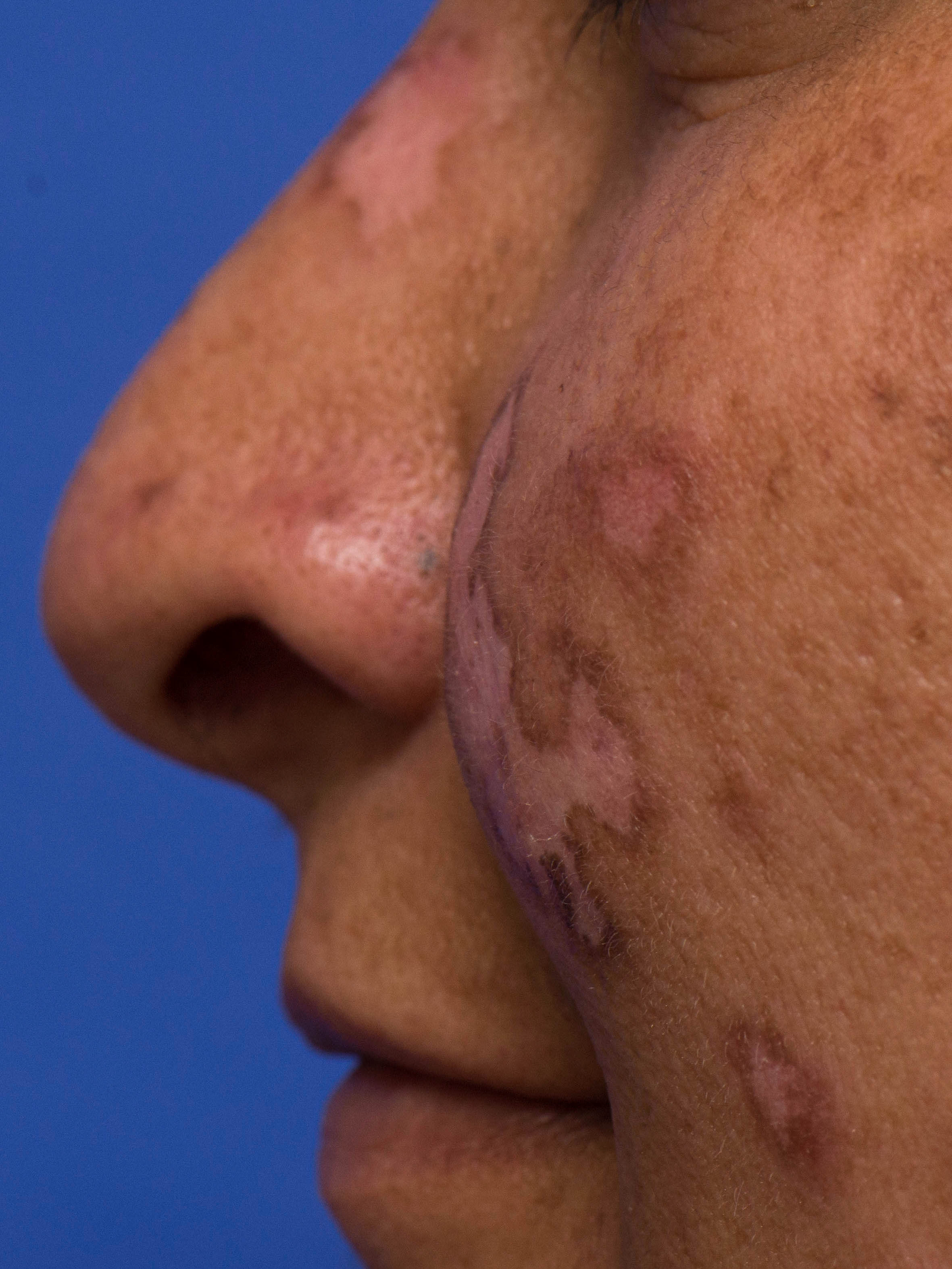
PROMISING THERAPEUTIC DEVELOPMENTS FOR CUTANEOUS LUPUS ERYTHEMATOSUS: INTERFERING WITH INTERFERON

By Warren R. Heymann, MD, FAAD
Aug. 17, 2022
Vol. 4, No. 33

Insights into the pathogenesis of lupus have encouraged trials of novel agents that may offer useful options for patients afflicted with CLE recalcitrant to existing therapies. According to Blake and Daniel: "LE is driven by dysfunction within the adaptive and innate immune system, beginning with a loss of self-tolerance in the adaptive immune system through the production of autoantibodies. These antibodies inappropriately react to the self-antigens present in cellular debris after apoptosis, resulting in activation and recruitment of T and B cells and production of immune complexes, which cause direct tissue injury. A number of proinflammatory signaling pathways are upregulated in patients with LE, which results in increased cytokine activity. The innate immune and complement systems are also pivotal for pathogen clearance, recognition of foreign antigens, and removal of apoptotic cells. Dysfunction in these two systems further drives LE manifestations." (3) Development of CLE likely commences in a pro-inflammatory epidermis, conditioned by excess type I interferon (IFN) production following ultraviolet light exposure. (4) The zinc finger transcription factors Ikaros and Aiolos are implicated in genetic predisposition to SLE. Ikaros induces development of B cells and plasmacytoid dendritic cells (pDCs, the major producers of type I interferon). Aiolos supports B-cell differentiation. (5)
Anifrolumab is a human, IgG1K monoclonal antibody that binds to the type 1 interferon receptor, thereby blocking type 1 interferon signaling. Anifrolimumab was approved by the FDA for SLE (though not lupus nephritis) in July 2021. (6) Blum et al identified 3 patients (Black women, aged 22 to 51 years) with CLE recalcitrant to standard therapies (variably — antimalarials, immunosuppressive agents, prednisone, rituximab, belimumab). All cases demonstrated significant improvement in disease appearance, cutaneous involvement, and symptomatology after treatment with 2 months of anifrolumab infusions added to their regimen. (7) Controlled trials are warranted to study to the potential of anifrolumab for CLE.

Sprow et al state: "IKZF1 and IKZF3 are susceptibility loci for SLE and encode the transcription factors Ikaros and Ailos. Cereblon is a molecule that forms part of a ubiquitin ligase complex which mediates the polyubiquitination and proteasome-dependent degradation of Ikaros and Ailos. Thalidomide and lenalidomide, drugs that have been used for off-label treatment of CLE, bind to cereblon resulting in increased destruction of Ikaros and Ailos, leading to decreased B cells, pDCs, and increased T regulatory cells." (6) In a 24-week, phase 2 trial involving patients with SLE, iberdomide (a cereblon modulator promoting degradation of the transcription factors Ikaros and Aiolos) resulted in a higher percentage of patients with a SLE Responder index-4 response than did placebo. CLASI-A was a secondary end point of this study; patients with CLE were more likely to have a reduction in their CLASI-A score with iberdomide. (5). Future studies are warranted in patients with CLE.
Although we have known that interferon plays a major role in the pathogenesis of lupus, our understanding has been incomplete. As our comprehension of interferon's precise pathogenic role becomes more refined, so will our therapeutic approaches that interfere with its effect.
Point to Remember: Advances in the knowledge of interferon's role in lupus pathogenesis are yielding therapeutic promise.
Our expert's viewpoint
Victoria P. Werth, MD, FAAD
Professor of Dermatology and Medicine, University of Pennsylvania School of Medicine
Chief of the Division of Dermatology, Philadelphia Veterans Administration Hospital
This is an exciting time for cutaneous lupus erythematosus (CLE). Recently published findings from three clinical trials show substantial progress in developing more targeted, efficacious, and better-tolerated medications for patients with CLE, particularly by inhibiting the production or action of type I interferons in affected skin. The NEJM just reported a phase 2 trial showing that litifilimab, a humanized monoclonal antibody against BDCA2, a receptor found on plasmacytoid dendritic cells (pDCs), significantly improved skin involvement in patients with CLE, with or without concurrent systemic LE (SLE). (8) Litifilimab causes internalization of the BDCA2 receptor and hence inhibition of local production of pro-inflammatory cytokines and chemokines, particularly type I interferons. A recent phase 1 study examining dadilimab, a humanized monoclonal antibody that depletes pDCs, showed inhibition of local cutaneous type I interferon responses — as well as significant improvements in CLE disease activity. (9) Anifrolumab is an antibody against the type I interferon receptor, thus blocking many type I interferons from binding and activating the interferon-α/β receptor (IFNAR). The FDA (USA) recently approved anifrolumab for SLE, based on a phase 3 trial that showed improved systemic symptoms, as well as improved lupus skin disease in patients with SLE. (10) Because SLE was an inclusion criterion, regulatory approval did not extend to patients with CLE who do not have concurrent SLE. The clinical trials of these three new agents present a consistent message — namely, that targeting type I interferons decreases cutaneous disease activity in many patients with skin findings of LE. All three of these trials used a validated disease severity tool, the cutaneous lupus erythematosus area and severity index (CLASI), which captures changes in skin that are meaningful to patients' quality of life, including total resolution and meaningful partial resolution. (11) Of note, the FDA typically allows regulatory approval for new medications that produce meaningful partial improvements in many diseases, yet the agency frequently mandates complete or nearly complete clearance of skin lesions before approval of new dermatologic medications. Such a regulatory approach might impede progress in CLE, in which partial improvements are often meaningful to patients. Moreover, there are currently only two FDA-approved medications for CLE, hydroxychloroquine and glucocorticoids, both of which have been in use since before 1962, when the FDA grandfathered them in without clinical trials. Issues for further study include heterogeneity of responses, since not all patients with CLE in the three recent trials improved, as well as the exact mechanisms by which therapeutic manipulations of plasmacytoid dendritic cells in two of the trials exerted beneficial effects. Beyond type I interferons and pDCs, there are many other targeted therapies in development for CLE. The findings of the NEJM paper and other novel studies will, we hope, allow development of medications with improved efficacy and fewer side effects in CLE than those currently used.
Dr. Werth had disclosed financial relationships with the following to the AAD at the time of publication: AbbVie, Akari Therapeutics, Amgen, arGEN-X, AstraZeneca, Bayer, Biogen, BMS, Celgene Corporation, Corbus Pharmaceuticals, Corcept Therapeutics, CSL, CSL Behring, Cugene, EMD Serono, Genentech, Inc., Gilead Sciences, GlaxoSmithKline, Idera Pharmaceuticals, Inc., Immune Pharmaceuticals, Immunotherapeutics Pharmaceuticals, Inc, Janssen Pharmaceuticals, Inc, Lilly ICOS LLC, Lupus Foundation of America, Medimmune, Neovacs, Octapharma, Pfizer Inc., Pincell, Principia Biopharma Inc, Q32 Bio Inc., Regeneron Pharmaceuticals, Inc., Resolve Therapeutics, Roche Laboratories, Stiefel a GSK company, Syntimmune, Inc., Timber Pharmaceuticals, UCB, UV Therapeutics, Vertex Pharmaceuticals Incorporated, Viela Bio. Full disclosure information is available at disclosures.aad.org.
Bitar C, Menge TD, Chan MP. Cutaneous manifestations of lupus erythematosus: a practical clinicopathological review for pathologists. Histopathology. 2022 Jan;80(1):233-250. doi: 10.1111/his.14440. PMID: 34197657.
Heymann WR. The criteria-dependent risk of systemic lupus erythematosus developing in patients with discoid lupus erythematosus. J Am Acad Dermatol. 2022 Jul 8:S0190-9622(22)02259-9. doi: 10.1016/j.jaad.2022.07.005. Epub ahead of print. PMID: 35810838.
Blake SC, Daniel BS. Cutaneous lupus erythematosus: A review of the literature. Int J Womens Dermatol. 2019 Jul 31;5(5):320-329. doi: 10.1016/j.ijwd.2019.07.004. PMID: 31909151; PMCID: PMC6938925.
Maz MP, Martens JWS, Hannoudi A, Reddy AL, Hile GA, Kahlenberg JM. Recent advances in cutaneous lupus. J Autoimmun. 2022 Jul 17:102865. doi: 10.1016/j.jaut.2022.102865. Epub ahead of print. PMID: 35858957.
Merrill JT, Werth VP, Furie R, van Vollenhoven R, Dörner T, Petronijevic M, Velasco J, Majdan M, Irazoque-Palazuelos F, Weiswasser M, Korish S, Ye Y, Gaudy A, Schafer PH, Liu Z, Agafonova N, Delev N. Phase 2 Trial of Iberdomide in Systemic Lupus Erythematosus. N Engl J Med. 2022 Mar 17;386(11):1034-1045. doi: 10.1056/NEJMoa2106535. PMID: 35294813.
Sprow G, Dan J, Merola JF, Werth VP. Emerging Therapies in Cutaneous Lupus Erythematosus. Front Med (Lausanne). 2022 Jul 11;9:968323. doi: 10.3389/fmed.2022.968323. PMID: 35899214; PMCID: PMC9313535.
Blum FR, Sampath AJ, Foulke GT. Anifrolumab for Treatment of Refractory Cutaneous Lupus Erythematosus. Clin Exp Dermatol. 2022 Jul 17. doi: 10.1111/ced.15335. Epub ahead of print. PMID: 35844070.
Werth VP, Furie RA, Romero-Diaz J, Navarra S, Kalunian K, van Vollenhoven RF, Nyberg F, Kaffenberger B, Sheikh SZ, Radunovic G, Huang X, Carroll H, Gaudreault F, Meyers A, Barbey C, Musselli C, Franchimont N, for the LILAC Study investigators. Trial of anti-BDCA2 antibody litifilimab for cutaneous lupus erythematosus. N Engl J Med. 287:321-331, 2022 (July 28). doi: 10.1056/NEJMoa2118024
Karnell JL, Wu Y, Mittereder N, Smith MA, Gunsior M, Yan L, Casey KA, Henault J, Riggs JM, Nicholson SM, Sanjuan MA, Vousden KA, Werth VP, Drappa J, Illei GG, Rees WA, Ratchford JN, for the VIB7734 Trial Investigators. Depleting plasmacytoid dendritic cells reduces local type I interferon responses and disease activity in patients with cutaneous lupus. Sci Transl Med. 2021;13:eabf8442. doi: 10.1126/scitranslmed.abf8442
Morand EF, Furie R, Tanaka Y, Bruce IN, Askanase AD, Richez C, et al. Trial of anifrolumab in active systemic lupus erythematosus. N Engl J Med. (2020) 382:211-21. doi: 10.1056/NEJMoa1912196
Chakka S, Krain RL, Ahmed S, Concha JSS, Feng R, Merrill JT, Werth VP. Evaluating change in disease activity needed to reflect meaningful improvement in quality of life for clinical trials in cutaneous lupus erythematosus. J Am Acad Dermatol 84(6):1562-1567, 2021.
All content found on Dermatology World Insights and Inquiries, including: text, images, video, audio, or other formats, were created for informational purposes only. The content represents the opinions of the authors and should not be interpreted as the official AAD position on any topic addressed. It is not intended to be a substitute for professional medical advice, diagnosis, or treatment.
Skin Care Physicians of Costa Rica
Clinica Victoria en San Pedro: 4000-1054
Momentum Escazu: 2101-9574
Please excuse the shortness of this message, as it has been sent from
a mobile device.
posted by dermatica at
August 25, 2022
![]()
![]()

0 Comments:
Post a Comment
Subscribe to Post Comments [Atom]
<< Home