Piel y Síndrome de Down
WHAT'S UP IN DOWN SYNDROME?

By Warren R. Heymann, MD, FAAD
October 13, 2021
Vol. 3, No. 41

DS is the most common chromosomal abnormality accounting for 8% of all registered cases. The incidence of Down syndrome is every 1 in 700 live births in the United States. DS is caused by the presence of all, or part of a third copy of chromosome 21. This extra genetic material is responsible for the classical facial characteristics, multiple malformations, intellectual disability, immune and endocrine dysfunction associated with DS. Although DS has a variable phenotype there are many common physical manifestations such as hypotonia, epicanthic folds, flat nasal bridge, single palmar crease, and sandal toe gap. This commentary will focus on some newer dermatological literature about DS. For a review of the cardiac, neurologic, gastrointestinal, endocrinologic, growth, respiratory, otolaryngologic, sleep, hematologic, immunologic, renal, and ophthalmologic comorbidities of DS, the reader is referred to Lagan et al for an excellent summary. (1)

Immunodysregulation in DS is complex, involving interferon-gamma (IFN-γ) hyperactivity and trisomy of the AIRE (autoimmune regulator) gene located on chromosome 21, reflected in the well-recognized association with autoimmune diseases such as alopecia areata, vitiligo, celiac disease, propensity to crusted scabies, multiple eruptive dermatofibromas, and type I diabetes. The interferon (IFN) response involves multiple cell types, causing hypersensitivity to IFN ligands, hyperactivation of downstream Janus kinase/signal transducer activator of transcription (JAK/STAT) signaling, and significant overexpression of IFN-stimulated genes. (2,3,4)
Rarer, but well-known associated dermatoses with DS include: Transient myeloproliferative disorder (TMD), elastosis perforans serpiginosa, syringomas, and milia-like calcinosis cutis. (4)
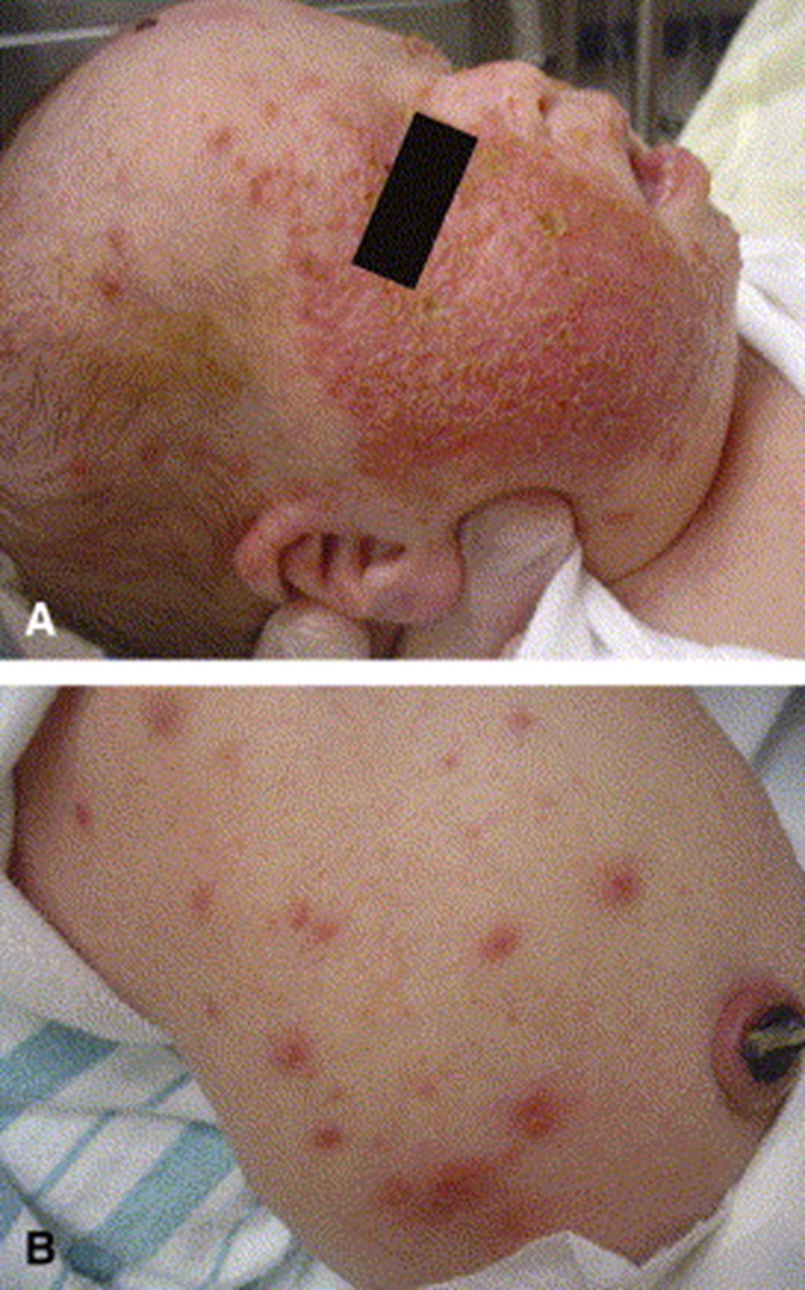
According to Brazzelli, "TMD is a spontaneously resolving clonal myeloid proliferation characterized by circulating megakaryoblasts in the peripheral blood that is restricted to neonates with Down syndrome (DS) or those with trisomy 21 mosaicism. Cutaneous manifestations of TMD are observed in only 5% of affected neonates and present as a diffuse eruption of erythematous, crusted papules, papulovesicles, and pustules, often with prominent and initial facial involvement." The dermatologic manifestations usually appear within the first 3 weeks of life, often starting on the face (cheeks), but may be distributed anywhere. Lesions last a few days and resolve without scarring. The leukemogenic source TMD is a somatic mutation of the X-linked GATA1 gene. Importantly, 20% of affected neonates ultimately develop acute leukemia later in life, typically with features of megakaryoblastic leukemia. (5) Patients with DS also have a twenty-fold increased risk of developing acute lymphoblastic leukemia (ALL). (1)
Regarding newer literature in DS, I found the following topics of interest.
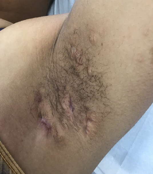
Lam et al performed a meta-analysis to study the association of hidradenitis suppurativa (HS) and DS. Twelve studies were included in this systematic review, with a total of 358 patients presenting with both HS and DS. Pooled analysis of mean differences between DS and non-DS participants presenting with HS found a significantly younger age of HS symptom onset for DS patients and a significantly increased likelihood of HS in DS patients. The authors suggest an association between HS and DS; this occurrence may be due to a combination of factors related to a propensity of follicular disorders in DS, immunodysregulation, and a trend toward obesity. (6)
The precise relationship of psoriasis with DS remains to be defined, although with the similarities in the Th1 pathway leading to production of interferon-gamma and tumor necrosis factor-alpha in both disorders, one would expect to see the two concurrently. Hedayati et al reported the case of annular psoriasis in a 17-year-old girl with DS. (7) Morita et al detail annular pustular psoriasis in an 8-year-old boy with DS following growth hormone therapy. (8) Senterre et al remind us to be careful with administration of biologics in patients with DS with presumed psoriasis. A 26-year-old woman was thought to have psoriasiform lesions for which the IL-23 inhibitor risankizumab was prescribed; unfortunately, the patient had crusted scabies which flared dramatically. She ultimately responded to topical permethrin and systemic ivermectin. (9)
Finally, Riga-Fede disease (traumatic granuloma of the tongue) which may have an alarming appearance mimicking malignancy, maybe due to the early onset and persistence of neonatal teeth, combined with uncontrolled [macroglossic, fissured] tongue movements in patients with DS. (10)
Point to Remember: Both common and rare disorders may be observed in patients with DS, but particular attention should be made on follicular, eczematous, immunologic disorders. Hidradenitis suppurativa is increasingly recognized in patients with DS. Multiorgan involvement mandates a multidisciplinary management approach.
Our expert's viewpoint
Jillian F. Rork, MD, FAAD
Assistant Professor of Dermatology
Dartmouth-Hitchcock Medical Center at Manchester and The Geisel School of Medicine
I have a phrase I like to teach about Down syndrome and skin: "Don't forget the hidden spots!" It is simplistic, but if you can recall this when examining a patient with Down syndrome, you will remember many of the common skin conditions that can be overlooked.

Make sure to look at the armpits, groin, and buttocks. Even in my young career, I have had numerous Down syndrome patients come in for different skin complaints only to find they have hidradenitis suppurativa (HS) or severe folliculitis on their buttocks. We, as dermatologists, know these can be painful, scarring skin conditions if untreated. While the literature is limited, it would appear that HS starts at a younger age in Down syndrome. We should be thinking about this diagnosis in school-aged and early adolescent patients.
Lastly, look at those feet! You could find hyperkeratosis that can impact how shoes fit and also dermatophyte infections. Onychomycosis can be found in younger patients with Down syndrome and should not just be considered in adults.
So, when seeing a Down syndrome patient, I would advocate for a complete skin examination. A cursory or focused examination may overlook skin conditions that could use our expertise.
Lagan N, Huggard D, Mc Grane F, Leahy TR, Franklin O, Roche E, Webb D, O' Marcaigh A, Cox D, El-Khuffash A, Greally P, Balfe J, Molloy EJ. Multiorgan involvement and management in children with Down syndrome. Acta Paediatr. 2020 Jun;109(6):1096-1111.
Rork JF, McCormack L, Lal K, Wiss K, Belazarian L. Dermatologic conditions in Down syndrome: A single-center retrospective chart review. Pediatr Dermatol. 2020 Sep;37(5):811-816.
Rachubinski AL, Estrada BE, Norris D, Dunnick CA, Boldrick JC, Espinosa JM. Janus kinase inhibition in Down syndrome: 2 cases of therapeutic benefit for alopecia areata. JAAD Case Rep. 2019 Apr 5;5(4):365-367.
Fölster-Holst R, Rohrer T, Jung AM. Dermatological aspects of the S2k guidelines on Down syndrome in childhood and adolescence. J Dtsch Dermatol Ges. 2018 Oct;16(10):1289-1295.
Brazzelli V, Segal A, Bernacca C, Tchich A, Bolcato V, Croci G, Mina T, Zecca M, Zanette S, Stronati M. Neonatal vesiculopustular eruption in Down syndrome and transient myeloproliferative disorder: A case report and review of the literature. Pediatr Dermatol. 2019 Sep;36(5):702-706.
Lam M, Lai C, Almuhanna N, Alhusayen R. Hidradenitis suppurativa and Down syndrome: A systematic review and meta-analysis. Pediatr Dermatol. 2020 Sep 6. doi: 10.1111/pde.14326. Epub ahead of print.
Hedayati B, Carley SK, Kraus CN, Smith J. Arcuate pink plaques in a female with Down syndrome. Int J Dermatol. 2020 Apr;59(4):e127-e128.
Morita A, Kawakami Y, Kaji T, Hirai Y, Miyake T, Takahashi M, Yamasaki O, Sugiura K, Morizane S. Pediatric-onset annular pustular psoriasis in a patient with Down syndrome. J Dermatol. 2019 Oct;46(10):e367-e368.
Senterre Y, Jouret G, Collins P, Nikkels AF. Risankizumab-Aggravated Crusted Scabies in a Patient with Down Syndrome. Dermatol Ther (Heidelb). 2020 Aug;10(4):829-834.
Polat Ekinci A, Kılıç S, Babuna Kobaner G. Early-onset and persistent traumatic granuloma of the tongue (Riga-Fede disease) associated with neonatal teeth and Down syndrome. J Eur Acad Dermatol Venereol. 2019 Mar;33(3):e131-e132.
Bull MJ; Committee on Genetics. Pediatrics. 2011;128(2):393-406
Tsou AY, Bulova P, Capone G, et al. Medical Care of Adults With Down Syndrome: A Clinical Guideline. JAMA.2020; 324(15):1543–1556.
Skin Care Physicians of Costa Rica
Clinica Victoria en San Pedro: 4000-1054
Momentum Escazu: 2101-9574
Please excuse the shortness of this message, as it has been sent from
a mobile device.
posted by dermatica at
October 21, 2021
![]()
![]()

0 Comments:
Post a Comment
Subscribe to Post Comments [Atom]
<< Home